Institute of Physiology, University of Zurich, Zurich, Switzerland.
National Centre of Competence in Research, Kidney.CH, Zurich, Switzerland.
Sci Data. 2023 Aug 3;10(1):510. doi: 10.1038/s41597-023-02407-5.
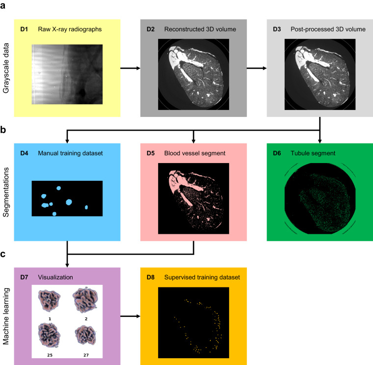
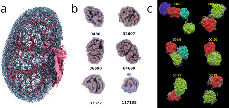
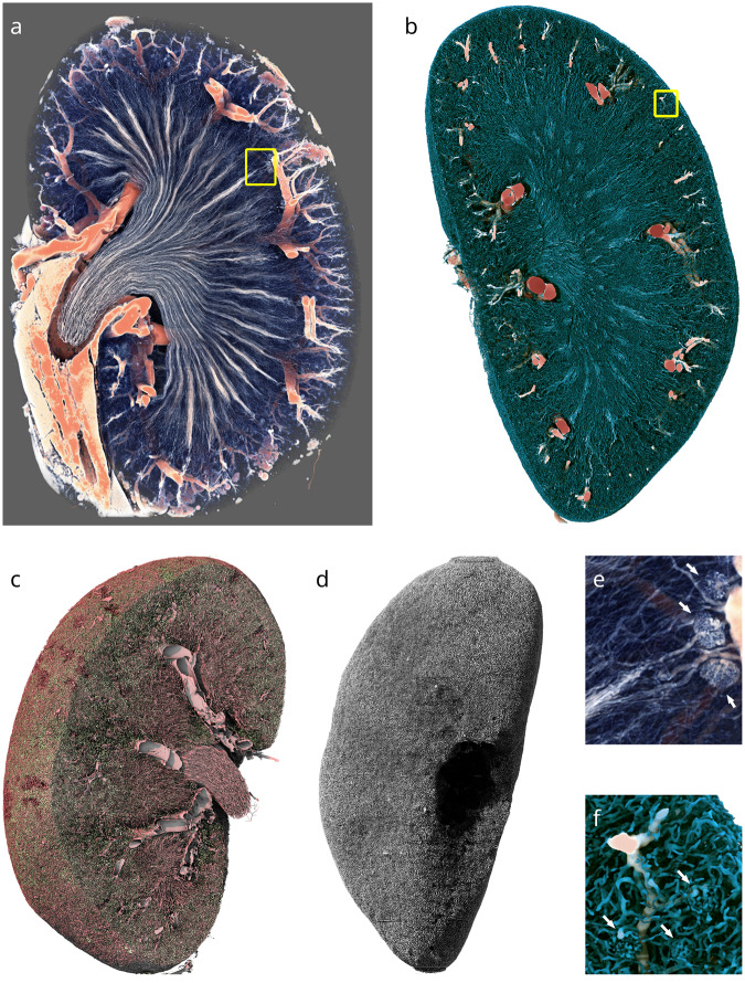
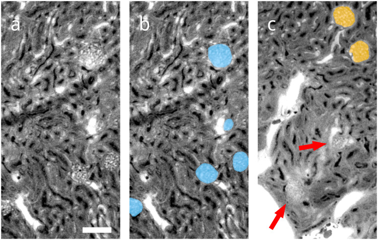
The performance of machine learning algorithms, when used for segmenting 3D biomedical images, does not reach the level expected based on results achieved with 2D photos. This may be explained by the comparative lack of high-volume, high-quality training datasets, which require state-of-the-art imaging facilities, domain experts for annotation and large computational and personal resources. The HR-Kidney dataset presented in this work bridges this gap by providing 1.7 TB of artefact-corrected synchrotron radiation-based X-ray phase-contrast microtomography images of whole mouse kidneys and validated segmentations of 33 729 glomeruli, which corresponds to a one to two orders of magnitude increase over currently available biomedical datasets. The image sets also contain the underlying raw data, threshold- and morphology-based semi-automatic segmentations of renal vasculature and uriniferous tubules, as well as true 3D manual annotations. We therewith provide a broad basis for the scientific community to build upon and expand in the fields of image processing, data augmentation and machine learning, in particular unsupervised and semi-supervised learning investigations, as well as transfer learning and generative adversarial networks.
机器学习算法在对 3D 生物医学图像进行分割时的性能并未达到基于 2D 照片所取得的结果预期的水平。这可能是由于缺乏大容量、高质量的训练数据集所致,而这些数据集需要最先进的成像设备、用于注释的领域专家以及大量的计算和个人资源。本文介绍的 HR-Kidney 数据集弥补了这一差距,它提供了 1.7TB 的经过校正的基于同步加速器辐射的全小鼠肾脏 X 射线相衬微断层摄影术图像,以及经过验证的 33729 个肾小球的分割结果,这与目前可用的生物医学数据集相比增加了一到两个数量级。这些图像集还包含基础原始数据、基于阈值和形态学的肾血管和尿细管半自动分割以及真实的 3D 手动注释。因此,我们为科学界提供了一个广泛的基础,可以在此基础上进行图像处理、数据增强和机器学习领域的研究,特别是无监督和半监督学习研究,以及迁移学习和生成对抗网络研究。