Department of Advanced Radiological Imaging, Kagoshima University Graduate School of Medical and Dental Sciences, 8-35-1 Sakuragaoka, Kagoshima, 890-8544, Japan.
Department of Radiology, Kagoshima University Graduate School of Medical and Dental Sciences, 8-35-1 Sakuragaoka, Kagoshima, 890-8544, Japan.
Cancer Imaging. 2023 Aug 8;23(1):75. doi: 10.1186/s40644-023-00595-2.
This study was designed to investigate the use of time-dependent diffusion magnetic resonance imaging (MRI) parameters in distinguishing between glioblastomas and brain metastases.
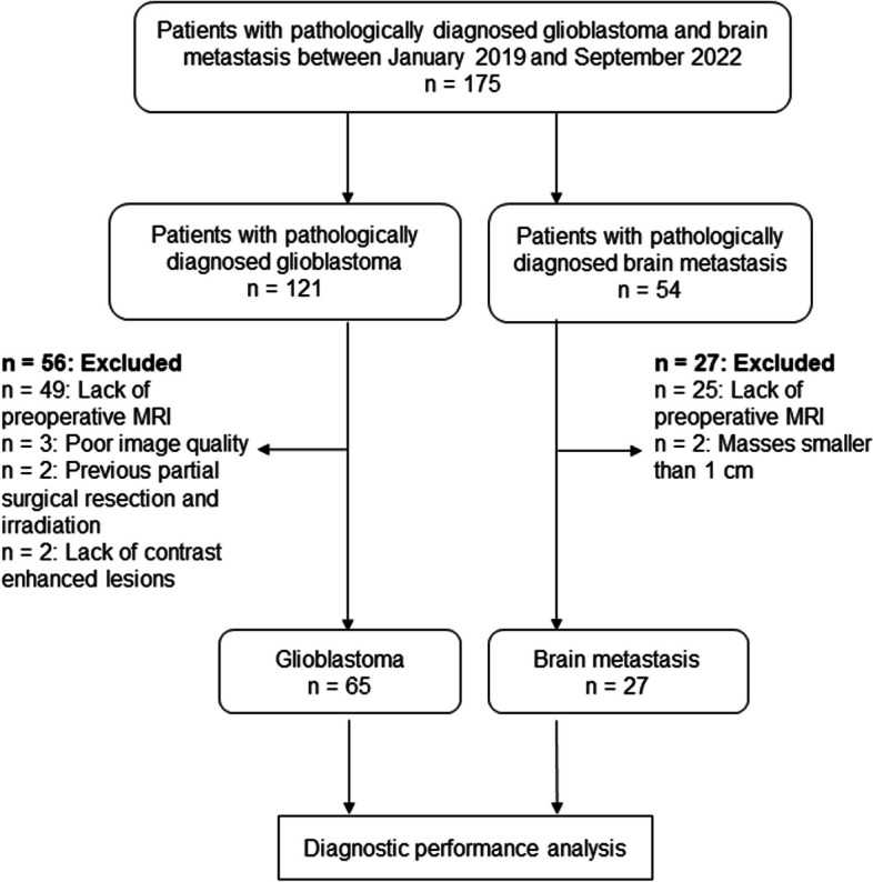
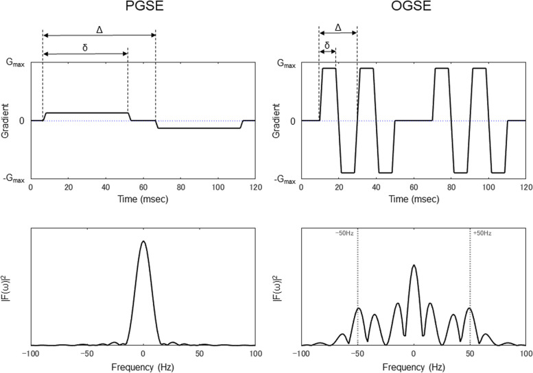
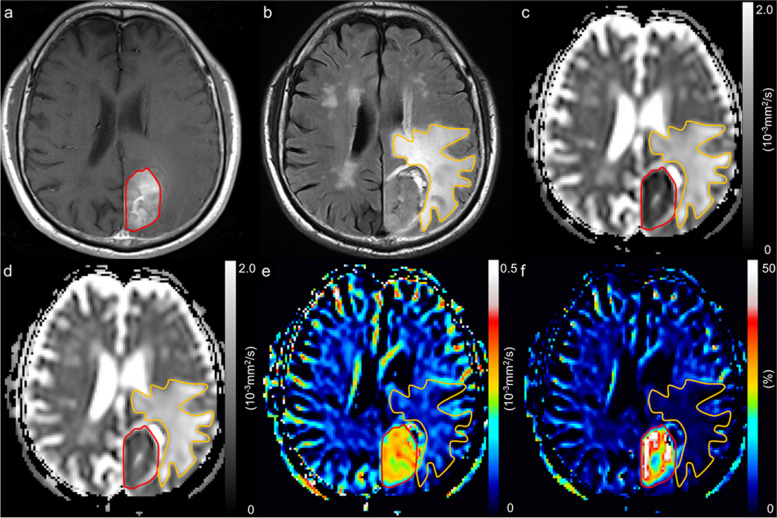
A retrospective study was conducted involving 65 patients with glioblastomas and 27 patients with metastases using a diffusion-weighted imaging sequence with oscillating gradient spin-echo (OGSE, 50 Hz) and a conventional pulsed gradient spin-echo (PGSE, 0 Hz) sequence. In addition to apparent diffusion coefficient (ADC) maps from two sequences (ADC and ADC), we generated maps of the ADC change (cADC): ADC - ADC and the relative ADC change (rcADC): (ADC - ADC)/ ADC × 100 (%).
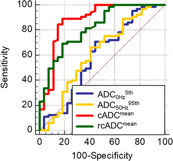
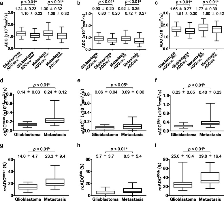
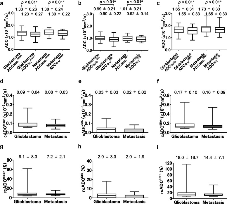
The mean and the fifth and 95th percentile values of each parameter in enhancing and peritumoral regions were compared between glioblastomas and metastases. The area under the receiver operating characteristic curve (AUC) values of the best discriminating indices were compared. In enhancing regions, none of the indices of ADC and ADC showed significant differences between metastases and glioblastomas. The mean cADC and rcADC values of metastases were significantly higher than those of glioblastomas (0.24 ± 0.12 × 10mm/s vs. 0.14 ± 0.03 × 10mm/s and 23.3 ± 9.4% vs. 14.0 ± 4.7%; all p < 0.01). In peritumoral regions, no significant difference in all ADC indices was observed between metastases and glioblastomas. The AUC values for the mean cADC (0.877) and rcADC (0.819) values in enhancing regions were significantly higher than those for ADC (0.595; all p < 0.001).
The time-dependent diffusion MRI parameters may be useful for differentiating brain metastases from glioblastomas.
本研究旨在探讨时间依赖性扩散磁共振成像(MRI)参数在鉴别胶质母细胞瘤和脑转移瘤中的应用。
回顾性研究纳入 65 例胶质母细胞瘤患者和 27 例转移瘤患者,使用扩散加权成像序列(带有振荡梯度回波自旋回波(OGSE,50 Hz)和常规脉冲梯度自旋回波(PGSE,0 Hz)序列)。除了两个序列(ADC 和 ADC)的表观扩散系数(ADC)图外,我们还生成了 ADC 变化图(cADC):ADC-ADC 和相对 ADC 变化图(rcADC):(ADC-ADC)/ADC×100(%)。
比较了增强区和瘤周区中每个参数的平均值和第 5 百分位和第 95 百分位值,比较了最佳鉴别指数的受试者工作特征曲线(ROC)下面积(AUC)值。在增强区,ADC 和 ADC 的各项指标在转移瘤和胶质母细胞瘤之间均无显著差异。转移瘤的平均 cADC 和 rcADC 值明显高于胶质母细胞瘤(0.24±0.12×10mm/s 与 0.14±0.03×10mm/s 和 23.3±9.4%与 14.0±4.7%;均 p<0.01)。在瘤周区,转移瘤和胶质母细胞瘤之间的所有 ADC 指标均无显著差异。增强区平均 cADC(0.877)和 rcADC(0.819)值的 AUC 值明显高于 ADC(0.595;均 p<0.001)。
时间依赖性扩散 MRI 参数可用于鉴别脑转移瘤和胶质母细胞瘤。