• 文献检索
  • 文档翻译
  • 深度研究
  • 学术资讯
  • Suppr Zotero 插件Zotero 插件
  • 邀请有礼
  • 套餐&价格
  • 历史记录
应用&插件
Suppr Zotero 插件Zotero 插件浏览器插件Mac 客户端Windows 客户端微信小程序
定价
高级版会员购买积分包购买API积分包
服务
文献检索文档翻译深度研究API 文档MCP 服务
关于我们
关于 Suppr公司介绍联系我们用户协议隐私条款
关注我们

Suppr 超能文献

核心技术专利:CN118964589B侵权必究
粤ICP备2023148730 号-1Suppr @ 2026

文献检索

告别复杂PubMed语法,用中文像聊天一样搜索,搜遍4000万医学文献。AI智能推荐,让科研检索更轻松。

立即免费搜索

文件翻译

保留排版,准确专业,支持PDF/Word/PPT等文件格式,支持 12+语言互译。

免费翻译文档

深度研究

AI帮你快速写综述,25分钟生成高质量综述,智能提取关键信息,辅助科研写作。

立即免费体验

基于MRI图像的多种深度学习模型鉴别胶质母细胞瘤与孤立性脑转移瘤:一项多中心研究

Multiple deep learning models based on MRI images in discriminating glioblastoma from solitary brain metastases: a multicentre study.

作者信息

Kong Chao, Yan Ding, Liu Kai, Yin Yong, Ma Changsheng

机构信息

Department of Graduate, Shandong First Medical University and Shandong Academy of Medical Sciences, Jinan, China.

Department of Radiation Physics, Shandong Cancer Hospital and Institute, Shandong First Medical University and Shandong Academy of Medical Sciences, Jinan, China.

出版信息

BMC Med Imaging. 2025 May 19;25(1):171. doi: 10.1186/s12880-025-01703-3.

DOI:10.1186/s12880-025-01703-3
PMID:40389875
原文链接:https://pmc.ncbi.nlm.nih.gov/articles/PMC12090387/
Abstract

OBJECTIVE

Development of a deep learning model for accurate preoperative identification of glioblastoma and solitary brain metastases by combining multi-centre and multi-sequence magnetic resonance images and comparison of the performance of different deep learning models.

METHODS

Clinical data and MR images of a total of 236 patients with pathologically confirmed glioblastoma and single brain metastases were retrospectively collected from January 2019 to May 2024 at Provincial Hospital of Shandong First Medical University, and the data were randomly divided into a training set and a test set according to the ratio of 8:2, in which the training set contained 197 cases and the test set contained 39 cases; the images were preprocessed and labeled with the tumor regions. The images were pre-processed and labeled with tumor regions, and different MRI sequences were input individually or in combination to train the deep learning model 3D ResNet-18, and the optimal sequence combinations were obtained by five-fold cross-validation enhancement of the data inputs and training of the deep learning models 3D Vision Transformer (3D Vit), 3D DenseNet, and 3D VGG; the working characteristic curves (ROCs) of subjects were plotted, and the area under the curve (AUC) was calculated. The area under the curve (AUC), accuracy, precision, recall and F1 score were used to evaluate the discriminative performance of the models. In addition, 48 patients with glioblastoma and single brain metastases from January 2020 to December 2022 were collected from the Affiliated Cancer Hospital of Shandong First Medical University as an external test set to compare the discriminative performance, robustness and generalization ability of the four deep learning models.

RESULTS

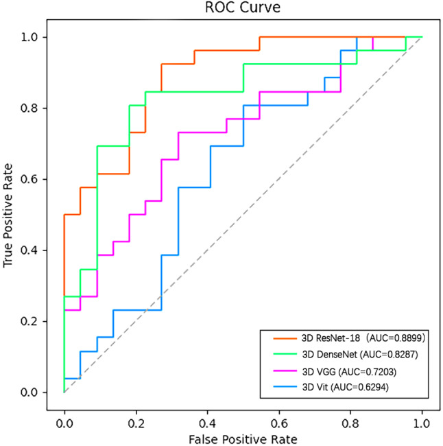
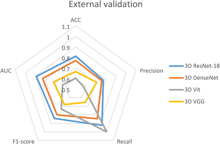
In the comparison of the discriminative effect of different MRI sequences, the three sequence combinations of T1-CE, T2, and T2-Flair gained discriminative effect, with the accuracy and AUC values of 0.8718 and 0.9305, respectively; after the four deep learning models were inputted into the aforementioned sequence combinations, the accuracy and AUC of the external validation of the 3D ResNet-18 model were 0.8125, respectively, 0.8899, all of which are the highest among all models.

CONCLUSIONS

A combination of multi-sequence MR images and a deep learning model can efficiently identify glioblastoma and solitary brain metastases preoperatively, and the deep learning model 3D ResNet-18 has the highest efficacy in identifying the two types of tumours.

摘要

目的

通过结合多中心和多序列磁共振图像,开发一种深度学习模型,用于术前准确识别胶质母细胞瘤和孤立性脑转移瘤,并比较不同深度学习模型的性能。

方法

回顾性收集2019年1月至2024年5月山东第一医科大学附属省立医院236例经病理证实的胶质母细胞瘤和单发脑转移瘤患者的临床资料和磁共振图像,并按照8:2的比例随机分为训练集和测试集,其中训练集包含197例,测试集包含39例;对图像进行预处理,并标记肿瘤区域。对图像进行预处理并标记肿瘤区域,将不同的磁共振成像序列单独或组合输入,训练深度学习模型3D ResNet-18,并通过对数据输入进行五折交叉验证增强以及训练深度学习模型3D视觉Transformer(3D Vit)、3D DenseNet和3D VGG获得最佳序列组合;绘制受试者工作特征曲线(ROC),并计算曲线下面积(AUC)。使用曲线下面积(AUC)、准确率、精确率、召回率和F1分数来评估模型的判别性能。此外,收集2020年1月至2022年12月山东第一医科大学附属肿瘤医院48例胶质母细胞瘤和单发脑转移瘤患者作为外部测试集,比较四种深度学习模型的判别性能、稳健性和泛化能力。

结果

在不同磁共振成像序列判别效果的比较中,T1-CE、T2和T2-Flair这三种序列组合获得了判别效果,准确率和AUC值分别为0.8718和0.9305;将四种深度学习模型输入上述序列组合后,3D ResNet-18模型外部验证的准确率和AUC分别为0.8125、0.8899,均为所有模型中最高。

结论

多序列磁共振图像与深度学习模型相结合能够有效术前识别胶质母细胞瘤和孤立性脑转移瘤,且深度学习模型3D ResNet-18在识别这两种肿瘤方面疗效最高。

https://cdn.ncbi.nlm.nih.gov/pmc/blobs/4660/12090387/151c5dec9964/12880_2025_1703_Fig6_HTML.jpg
https://cdn.ncbi.nlm.nih.gov/pmc/blobs/4660/12090387/f02096125198/12880_2025_1703_Fig1_HTML.jpg
https://cdn.ncbi.nlm.nih.gov/pmc/blobs/4660/12090387/16a7714c921d/12880_2025_1703_Fig2_HTML.jpg
https://cdn.ncbi.nlm.nih.gov/pmc/blobs/4660/12090387/2bce2a1e42b2/12880_2025_1703_Fig3_HTML.jpg
https://cdn.ncbi.nlm.nih.gov/pmc/blobs/4660/12090387/e9f46a20783f/12880_2025_1703_Fig4_HTML.jpg
https://cdn.ncbi.nlm.nih.gov/pmc/blobs/4660/12090387/68009eea9cf2/12880_2025_1703_Fig5_HTML.jpg
https://cdn.ncbi.nlm.nih.gov/pmc/blobs/4660/12090387/151c5dec9964/12880_2025_1703_Fig6_HTML.jpg

相似文献

1
Multiple deep learning models based on MRI images in discriminating glioblastoma from solitary brain metastases: a multicentre study.基于MRI图像的多种深度学习模型鉴别胶质母细胞瘤与孤立性脑转移瘤:一项多中心研究
BMC Med Imaging. 2025 May 19;25(1):171. doi: 10.1186/s12880-025-01703-3.
2
Development and Validation of a Deep Learning-Based Model to Distinguish Glioblastoma from Solitary Brain Metastasis Using Conventional MR Images.基于深度学习的模型在利用常规磁共振成像区分脑胶质母细胞瘤和单发脑转移瘤中的建立与验证。
AJNR Am J Neuroradiol. 2021 May;42(5):838-844. doi: 10.3174/ajnr.A7003. Epub 2021 Mar 18.
3
Development and validation of a multi-modality fusion deep learning model for differentiating glioblastoma from solitary brain metastases.开发和验证一种多模态融合深度学习模型,用于区分胶质母细胞瘤和单发脑转移瘤。
Zhong Nan Da Xue Xue Bao Yi Xue Ban. 2024 Jan 28;49(1):58-67. doi: 10.11817/j.issn.1672-7347.2024.230248.
4
Glioblastoma and Solitary Brain Metastasis: Differentiation by Integrating Demographic-MRI and Deep-Learning Radiomics Signatures.胶质母细胞瘤和单发脑转移瘤:通过整合人口统计学 MRI 和深度学习放射组学特征进行区分。
J Magn Reson Imaging. 2024 Sep;60(3):909-920. doi: 10.1002/jmri.29123. Epub 2023 Nov 13.
5
Differentiation Between Glioblastoma and Metastatic Disease on Conventional MRI Imaging Using 3D-Convolutional Neural Networks: Model Development and Validation.利用三维卷积神经网络在常规 MRI 成像上区分脑胶质母细胞瘤和转移性疾病:模型的建立与验证。
Acad Radiol. 2024 May;31(5):2041-2049. doi: 10.1016/j.acra.2023.10.044. Epub 2023 Nov 17.
6
Texture Feature Differentiation of Glioblastoma and Solitary Brain Metastases Based on Tumor and Tumor-brain Interface.基于肿瘤及肿瘤-脑界面的胶质母细胞瘤与孤立性脑转移瘤的纹理特征鉴别
Acad Radiol. 2025 Jan;32(1):400-410. doi: 10.1016/j.acra.2024.08.025. Epub 2024 Aug 31.
7
Deep learning enables automatic detection and segmentation of brain metastases on multisequence MRI.深度学习可实现多序列 MRI 上脑转移瘤的自动检测和分割。
J Magn Reson Imaging. 2020 Jan;51(1):175-182. doi: 10.1002/jmri.26766. Epub 2019 May 2.
8
Discrimination Between Glioblastoma and Solitary Brain Metastasis Using Conventional MRI and Diffusion-Weighted Imaging Based on a Deep Learning Algorithm.基于深度学习算法的常规 MRI 和弥散加权成像在胶质母细胞瘤与单发脑转移瘤鉴别诊断中的应用。
J Digit Imaging. 2023 Aug;36(4):1480-1488. doi: 10.1007/s10278-023-00838-5. Epub 2023 May 8.
9
Differentiation between glioblastoma, brain metastasis and subtypes using radiomics analysis.使用放射组学分析对胶质母细胞瘤、脑转移瘤和亚型进行区分。
J Magn Reson Imaging. 2019 Aug;50(2):519-528. doi: 10.1002/jmri.26643. Epub 2019 Jan 11.
10
Ensemble learning-based radiomics model for discriminating brain metastasis from glioblastoma.基于集成学习的用于鉴别胶质母细胞瘤脑转移的放射组学模型。
Eur J Radiol. 2025 Feb;183:111900. doi: 10.1016/j.ejrad.2024.111900. Epub 2024 Dec 24.

本文引用的文献

1
NUT carcinoma of the head and neck: A case report and literature review.头颈部NUT癌:一例报告及文献综述。
Precis Radiat Oncol. 2024 Sep 1;8(3):138-144. doi: 10.1002/pro6.1237. eCollection 2024 Sep.
2
Automated multi-class MRI brain tumor classification and segmentation using deformable attention and saliency mapping.使用可变形注意力和显著性映射的自动多类别MRI脑肿瘤分类与分割
Sci Rep. 2025 Mar 8;15(1):8114. doi: 10.1038/s41598-025-92776-1.
3
An assessment of breast cancer HER2, ER, and PR expressions based on mammography using deep learning with convolutional neural networks.
基于乳腺钼靶摄影,利用卷积神经网络深度学习对乳腺癌HER2、ER和PR表达进行评估。
Sci Rep. 2025 Feb 9;15(1):4826. doi: 10.1038/s41598-024-83597-9.
4
Brain tumor detection and segmentation using deep learning.使用深度学习进行脑肿瘤检测与分割
MAGMA. 2025 Feb;38(1):13-22. doi: 10.1007/s10334-024-01203-5. Epub 2024 Sep 4.
5
A comprehensive survey on deep active learning in medical image analysis.医学图像分析中深度主动学习的全面综述。
Med Image Anal. 2024 Jul;95:103201. doi: 10.1016/j.media.2024.103201. Epub 2024 May 21.
6
MRI radiomics in head and neck cancer from reproducibility to combined approaches.头颈部癌中的MRI影像组学:从可重复性到联合方法
Sci Rep. 2024 Apr 24;14(1):9451. doi: 10.1038/s41598-024-60009-6.
7
Editorial for "Glioblastoma and Solitary Brain Metastasis: Differentiation by Integrating Demographic-MRI and Deep-Learning Radiomics Signatures".《胶质母细胞瘤与孤立性脑转移瘤:通过整合人口统计学 - MRI 及深度学习影像组学特征进行鉴别》的社论
J Magn Reson Imaging. 2024 Sep;60(3):921-922. doi: 10.1002/jmri.29126. Epub 2023 Nov 16.
8
Distinguishing Tumor Cell Infiltration and Vasogenic Edema in the Peritumoral Region of Glioblastoma at the Voxel Level via Conventional MRI Sequences.通过常规 MRI 序列在脑胶质母细胞瘤瘤周区域的体素水平区分肿瘤细胞浸润和血管源性水肿。
Acad Radiol. 2024 Mar;31(3):1082-1090. doi: 10.1016/j.acra.2023.08.008. Epub 2023 Sep 14.
9
Differentiating brain metastasis from glioblastoma by time-dependent diffusion MRI.基于时变弥散 MRI 鉴别脑转移瘤与胶质母细胞瘤。
Cancer Imaging. 2023 Aug 8;23(1):75. doi: 10.1186/s40644-023-00595-2.
10
ResNet and its application to medical image processing: Research progress and challenges.ResNet 及其在医学图像处理中的应用:研究进展与挑战。
Comput Methods Programs Biomed. 2023 Oct;240:107660. doi: 10.1016/j.cmpb.2023.107660. Epub 2023 Jun 8.