• 文献检索
  • 文档翻译
  • 深度研究
  • 学术资讯
  • Suppr Zotero 插件Zotero 插件
  • 邀请有礼
  • 套餐&价格
  • 历史记录
应用&插件
Suppr Zotero 插件Zotero 插件浏览器插件Mac 客户端Windows 客户端微信小程序
定价
高级版会员购买积分包购买API积分包
服务
文献检索文档翻译深度研究API 文档MCP 服务
关于我们
关于 Suppr公司介绍联系我们用户协议隐私条款
关注我们

Suppr 超能文献

核心技术专利:CN118964589B侵权必究
粤ICP备2023148730 号-1Suppr @ 2026

文献检索

告别复杂PubMed语法,用中文像聊天一样搜索,搜遍4000万医学文献。AI智能推荐,让科研检索更轻松。

立即免费搜索

文件翻译

保留排版,准确专业,支持PDF/Word/PPT等文件格式,支持 12+语言互译。

免费翻译文档

深度研究

AI帮你快速写综述,25分钟生成高质量综述,智能提取关键信息,辅助科研写作。

立即免费体验

通过冷冻电子断层扫描揭示了顶复门寄生虫滑行运动中肌动蛋白丝的起源和排列。

Origin and arrangement of actin filaments for gliding motility in apicomplexan parasites revealed by cryo-electron tomography.

机构信息

Department of Biochemistry and Biophysics, Perelman School of Medicine, University of Pennsylvania, Philadelphia, PA, USA.

Institute of Structural Biology, Perelman School of Medicine, University of Pennsylvania, Philadelphia, PA, USA.

出版信息

Nat Commun. 2023 Aug 9;14(1):4800. doi: 10.1038/s41467-023-40520-6.

DOI:10.1038/s41467-023-40520-6
PMID:37558667
原文链接:https://pmc.ncbi.nlm.nih.gov/articles/PMC10412601/
Abstract

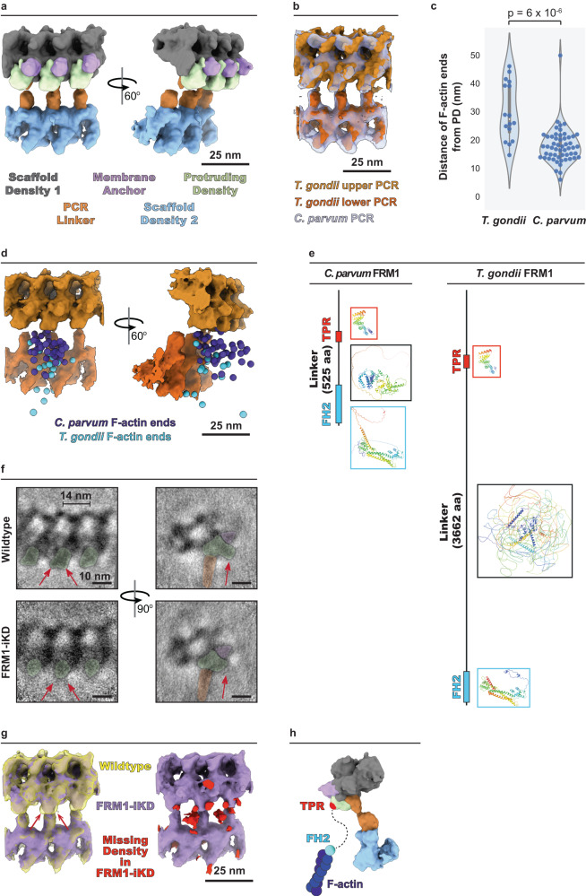
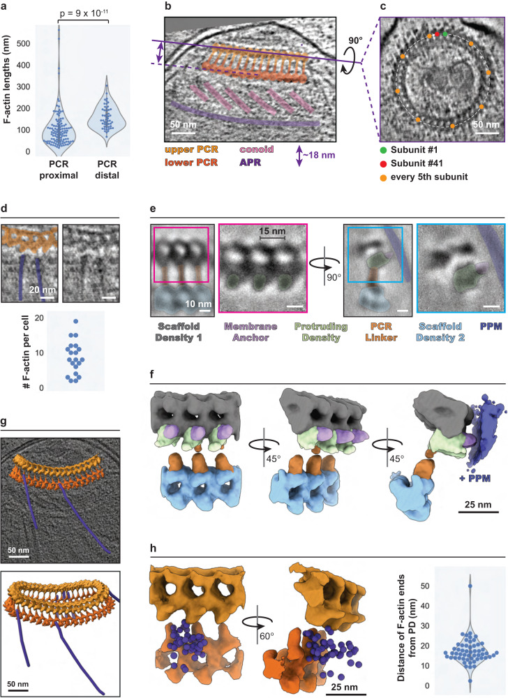
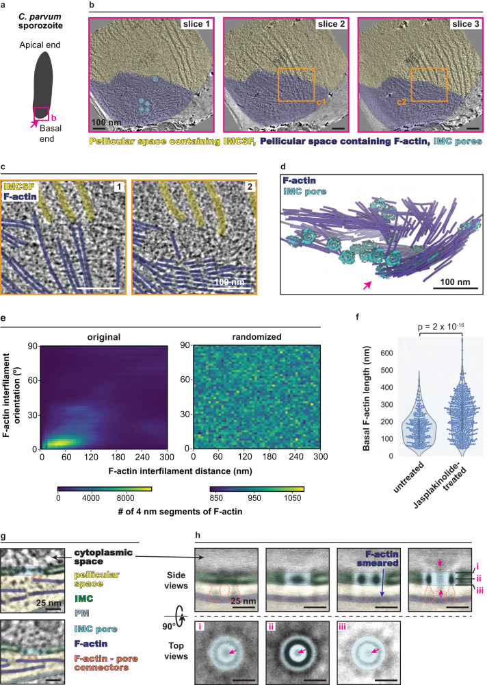
The phylum Apicomplexa comprises important eukaryotic parasites that invade host tissues and cells using a unique mechanism of gliding motility. Gliding is powered by actomyosin motors that translocate host-attached surface adhesins along the parasite cell body. Actin filaments (F-actin) generated by Formin1 play a central role in this critical parasitic activity. However, their subcellular origin, path and ultrastructural arrangement are poorly understood. Here we used cryo-electron tomography to image motile Cryptosporidium parvum sporozoites and reveal the cellular architecture of F-actin at nanometer-scale resolution. We demonstrate that F-actin nucleates at the apically positioned preconoidal rings and is channeled into the pellicular space between the parasite plasma membrane and the inner membrane complex in a conoid extrusion-dependent manner. Within the pellicular space, filaments on the inner membrane complex surface appear to guide the apico-basal flux of F-actin. F-actin concordantly accumulates at the basal end of the parasite. Finally, analyzing a Formin1-depleted Toxoplasma gondii mutant pinpoints the upper preconoidal ring as the conserved nucleation hub for F-actin in Cryptosporidium and Toxoplasma. Together, we provide an ultrastructural model for the life cycle of F-actin for apicomplexan gliding motility.

摘要

肉足鞭毛门包含重要的真核寄生虫,它们利用独特的滑行运动机制入侵宿主组织和细胞。滑行运动由肌动球蛋白马达驱动,该马达沿着寄生虫细胞本体将附着在宿主上的表面黏附素向前输送。Formin1 产生的肌动蛋白丝 (F-actin) 在这一关键的寄生活动中发挥着核心作用。然而,它们的亚细胞起源、路径和超微结构排列仍知之甚少。在这里,我们使用冷冻电子断层扫描来对运动中的微小隐孢子虫孢子虫进行成像,并以纳米级分辨率揭示 F-actin 的细胞结构。我们证明 F-actin 在顶端定位的 preconoidal 环上起始,并以依赖于锥体挤出的方式被引导到寄生虫质膜和内膜复合物之间的膜下空间中。在膜下空间中,内膜复合物表面上的细丝似乎引导 F-actin 的顶-基极流。F-actin 一致地在寄生虫的基底末端积累。最后,分析 Formin1 缺失的刚地弓形虫突变体指出,上预锥环是隐孢子虫和刚地弓形虫中 F-actin 的保守起始枢纽。总的来说,我们为肉足鞭毛门的滑行运动提供了 F-actin 生命周期的超微结构模型。

https://cdn.ncbi.nlm.nih.gov/pmc/blobs/7fed/10412601/6d9b810f4ab1/41467_2023_40520_Fig6_HTML.jpg
https://cdn.ncbi.nlm.nih.gov/pmc/blobs/7fed/10412601/e1ec994dab77/41467_2023_40520_Fig1_HTML.jpg
https://cdn.ncbi.nlm.nih.gov/pmc/blobs/7fed/10412601/1898b6cc1920/41467_2023_40520_Fig2_HTML.jpg
https://cdn.ncbi.nlm.nih.gov/pmc/blobs/7fed/10412601/1b402532975a/41467_2023_40520_Fig3_HTML.jpg
https://cdn.ncbi.nlm.nih.gov/pmc/blobs/7fed/10412601/9eff08a15bac/41467_2023_40520_Fig4_HTML.jpg
https://cdn.ncbi.nlm.nih.gov/pmc/blobs/7fed/10412601/1779d8b813d1/41467_2023_40520_Fig5_HTML.jpg
https://cdn.ncbi.nlm.nih.gov/pmc/blobs/7fed/10412601/6d9b810f4ab1/41467_2023_40520_Fig6_HTML.jpg

相似文献

1
Origin and arrangement of actin filaments for gliding motility in apicomplexan parasites revealed by cryo-electron tomography.通过冷冻电子断层扫描揭示了顶复门寄生虫滑行运动中肌动蛋白丝的起源和排列。
Nat Commun. 2023 Aug 9;14(1):4800. doi: 10.1038/s41467-023-40520-6.
2
Conoid extrusion regulates glideosome assembly to control motility and invasion in Apicomplexa.锥体外排调节滑行体组装以控制顶复门动物的运动和侵袭。
Nat Microbiol. 2022 Nov;7(11):1777-1790. doi: 10.1038/s41564-022-01212-x. Epub 2022 Sep 15.
3
Evolutionarily divergent, unstable filamentous actin is essential for gliding motility in apicomplexan parasites.进化上不同,不稳定的丝状肌动蛋白对于顶复门寄生虫的滑行运动是必不可少的。
PLoS Pathog. 2011 Oct;7(10):e1002280. doi: 10.1371/journal.ppat.1002280. Epub 2011 Oct 6.
4
Surface attachment, promoted by the actomyosin system of Toxoplasma gondii is important for efficient gliding motility and invasion.由刚地弓形虫的肌动球蛋白系统促进的表面附着对于有效的滑行运动和入侵很重要。
BMC Biol. 2017 Jan 18;15(1):1. doi: 10.1186/s12915-016-0343-5.
5
Unusual kinetic and structural properties control rapid assembly and turnover of actin in the parasite Toxoplasma gondii.异常的动力学和结构特性控制着寄生虫弓形虫中肌动蛋白的快速组装和周转。
Mol Biol Cell. 2006 Feb;17(2):895-906. doi: 10.1091/mbc.e05-06-0512. Epub 2005 Nov 30.
6
Actin filament polymerization regulates gliding motility by apicomplexan parasites.肌动蛋白丝聚合调节顶复门寄生虫的滑行运动。
Mol Biol Cell. 2003 Feb;14(2):396-406. doi: 10.1091/mbc.e02-08-0458.
7
Actin depolymerizing factor controls actin turnover and gliding motility in Toxoplasma gondii.肌动蛋白解聚因子控制刚地弓形虫中的肌动蛋白周转和滑行运动。
Mol Biol Cell. 2011 Apr 15;22(8):1290-9. doi: 10.1091/mbc.E10-12-0939. Epub 2011 Feb 23.
8
Structural and regulatory insights into the glideosome-associated connector from .对. 滑行体相关连接器的结构和调节见解
Elife. 2023 Apr 4;12:e86049. doi: 10.7554/eLife.86049.
9
In situ ultrastructures of two evolutionarily distant apicomplexan rhoptry secretion systems.两种进化上相距甚远的顶复门微线体分泌系统的原位超微结构。
Nat Commun. 2021 Aug 17;12(1):4983. doi: 10.1038/s41467-021-25309-9.
10
An Apicomplexan Actin-Binding Protein Serves as a Connector and Lipid Sensor to Coordinate Motility and Invasion.一种质体锥虫肌动蛋白结合蛋白作为连接蛋白和脂质传感器,协调运动和入侵。
Cell Host Microbe. 2016 Dec 14;20(6):731-743. doi: 10.1016/j.chom.2016.10.020.

引用本文的文献

1
New dimensions in acidocalcisome research: the potential of cryo-EM to uncover novel aspects of protozoan parasite physiology.酸性钙小体研究的新维度:冷冻电镜揭示原生动物寄生虫生理学新方面的潜力
mBio. 2025 May 14;16(5):e0166224. doi: 10.1128/mbio.01662-24. Epub 2025 Apr 8.
2
Molecular architecture of glideosome and nuclear F-actin in Plasmodium falciparum.恶性疟原虫中滑行体和细胞核丝状肌动蛋白的分子结构
EMBO Rep. 2025 Apr;26(8):1984-1996. doi: 10.1038/s44319-025-00415-7. Epub 2025 Mar 24.
3
Cryo-ET reveals the in situ architecture of the polar tube invasion apparatus from microsporidian parasites.

本文引用的文献

1
Cryogenic electron tomography reveals novel structures in the apical complex of .低温电子断层成像揭示了 顶端复合物的新结构。
mBio. 2024 Apr 10;15(4):e0286423. doi: 10.1128/mbio.02864-23. Epub 2024 Mar 8.
2
Nanoscale details of mitochondrial constriction revealed by cryoelectron tomography.冷冻电镜断层成像术揭示的线粒体缢缩的纳米级细节。
Biophys J. 2023 Sep 19;122(18):3768-3782. doi: 10.1016/j.bpj.2023.07.030. Epub 2023 Aug 1.
3
Cryo-tomography reveals rigid-body motion and organization of apicomplexan invasion machinery.
冷冻电子断层扫描揭示了微孢子虫寄生虫极管侵入装置的原位结构。
Proc Natl Acad Sci U S A. 2025 Mar 18;122(11):e2415233122. doi: 10.1073/pnas.2415233122. Epub 2025 Mar 11.
4
The big chill: Growth of structural biology with cryo-electron tomography.大变革:冷冻电子断层扫描技术助力结构生物学发展
QRB Discov. 2024 Dec 13;5:e10. doi: 10.1017/qrd.2024.10. eCollection 2024.
5
Emergent actin flows explain distinct modes of gliding motility.紧急肌动蛋白流解释了不同的滑行运动模式。
Nat Phys. 2024;20(12):1989-1996. doi: 10.1038/s41567-024-02652-4. Epub 2024 Oct 8.
6
Architecture of the apical polar ring and its role in gliding motility and invasion.顶端极环的结构及其在滑行运动和侵袭中的作用。
Proc Natl Acad Sci U S A. 2024 Nov 12;121(46):e2416602121. doi: 10.1073/pnas.2416602121. Epub 2024 Nov 8.
7
Submicrometre spatiotemporal characterization of the Toxoplasma adhesion strategy for gliding motility.微纳尺度时空解析弓形虫滑行运动的黏附策略。
Nat Microbiol. 2024 Dec;9(12):3148-3164. doi: 10.1038/s41564-024-01818-3. Epub 2024 Nov 4.
8
γ-tubulin complex controls the nucleation of tubulin-based structures in Apicomplexa.γ-微管蛋白复合物控制锥体虫类基于微管的结构的成核。
Mol Biol Cell. 2024 Sep 1;35(9):ar121. doi: 10.1091/mbc.E24-03-0100. Epub 2024 Jul 24.
9
Contribution of microscopy to a better understanding of the anatomy of pathogenic protists.显微镜检查对更好地了解致病原生动物的解剖结构的贡献。
Proc Natl Acad Sci U S A. 2024 Apr 23;121(17):e2321515121. doi: 10.1073/pnas.2321515121. Epub 2024 Apr 15.
10
Cryogenic electron tomography reveals novel structures in the apical complex of .低温电子断层成像揭示了 顶端复合物的新结构。
mBio. 2024 Apr 10;15(4):e0286423. doi: 10.1128/mbio.02864-23. Epub 2024 Mar 8.
低温断层成像揭示了质体入侵机械的刚体运动和组织。
Nat Commun. 2023 Mar 30;14(1):1775. doi: 10.1038/s41467-023-37327-w.
4
Cryo-electron tomography with mixed-scale dense neural networks reveals key steps in deployment of invasion machinery.结合混合尺度密集神经网络的冷冻电子断层扫描揭示了侵袭机制部署中的关键步骤。
PNAS Nexus. 2022 Sep 4;1(4):pgac183. doi: 10.1093/pnasnexus/pgac183. eCollection 2022 Sep.
5
Conoid extrusion regulates glideosome assembly to control motility and invasion in Apicomplexa.锥体外排调节滑行体组装以控制顶复门动物的运动和侵袭。
Nat Microbiol. 2022 Nov;7(11):1777-1790. doi: 10.1038/s41564-022-01212-x. Epub 2022 Sep 15.
6
Divergent Plasmodium actin residues are essential for filament localization, mosquito salivary gland invasion and malaria transmission.不同的疟原虫肌动蛋白残基对于丝状体的定位、蚊子唾液腺入侵和疟疾传播是必不可少的。
PLoS Pathog. 2022 Aug 23;18(8):e1010779. doi: 10.1371/journal.ppat.1010779. eCollection 2022 Aug.
7
Rhoptry secretion system structure and priming in Plasmodium falciparum revealed using in situ cryo-electron tomography.利用原位冷冻电子断层成像技术揭示恶性疟原虫裂殖子分泌系统的结构和启动机制。
Nat Microbiol. 2022 Aug;7(8):1230-1238. doi: 10.1038/s41564-022-01171-3. Epub 2022 Jul 11.
8
The BCC7 Protein Contributes to the Basal Pole by Interfacing between the MyoC Motor and the IMC Membrane Network.BCC7 蛋白通过与 MyoC 马达和 IMC 膜网络之间的相互作用,有助于基极。
Int J Mol Sci. 2022 May 26;23(11):5995. doi: 10.3390/ijms23115995.
9
's Basal Complex: The Other Apicomplexan Business End Is Multifunctional.'s Basal Complex:其他顶复门的“末端”是多功能的。
Front Cell Infect Microbiol. 2022 Apr 29;12:882166. doi: 10.3389/fcimb.2022.882166. eCollection 2022.
10
Cofilactin filaments regulate filopodial structure and dynamics in neuronal growth cones.丝状肌动蛋白丝调节神经元生长锥中的丝状伪足结构和动力学。
Nat Commun. 2022 May 4;13(1):2439. doi: 10.1038/s41467-022-30116-x.