Hueschen Christina L, Segev-Zarko Li-Av, Chen Jian-Hua, LeGros Mark A, Larabell Carolyn A, Boothroyd John C, Phillips Rob, Dunn Alexander R
Dept. of Chemical Engineering, Stanford University, Palo Alto, CA USA.
Present Address: Dept. of Cell and Developmental Biology, University of California San Diego, La Jolla, CA USA.
Nat Phys. 2024;20(12):1989-1996. doi: 10.1038/s41567-024-02652-4. Epub 2024 Oct 8.
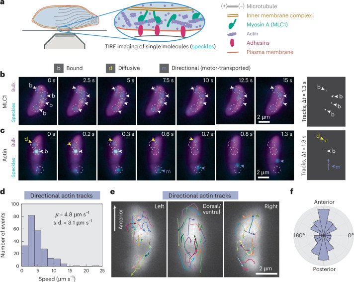
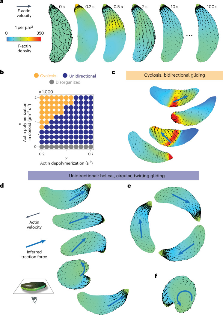
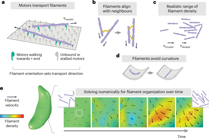
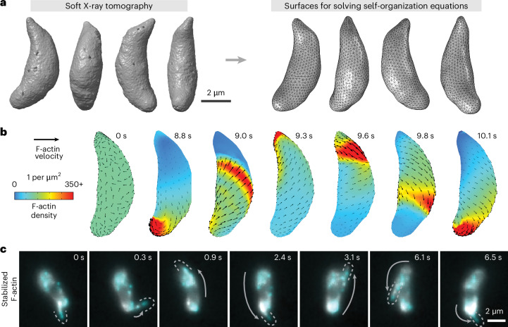
During host infection, and related unicellular parasites move using gliding, which differs fundamentally from other known mechanisms of eukaryotic cell motility. Gliding is thought to be powered by a thin layer of flowing filamentous (F)-actin sandwiched between the plasma membrane and a myosin-covered inner membrane complex. How this surface actin layer drives the various gliding modes observed in experiments-helical, circular, twirling and patch, pendulum or rolling-is unclear. Here we suggest that F-actin flows arise through self-organization and develop a continuum model of emergent F-actin flow within the confines provided by geometry. In the presence of F-actin turnover, our model predicts the emergence of a steady-state mode in which actin transport is largely directed rearward. Removing F-actin turnover leads to actin patches that recirculate up and down the cell, which we observe experimentally for drug-stabilized actin bundles in live parasites. These distinct self-organized actin states can account for observed gliding modes, illustrating how different forms of gliding motility can emerge as an intrinsic consequence of the self-organizing properties of F-actin flow in a confined geometry.
在宿主感染期间,相关单细胞寄生虫通过滑行移动,这与真核细胞运动的其他已知机制有根本区别。滑行被认为是由夹在质膜和肌球蛋白覆盖的内膜复合体之间的一层薄薄的流动丝状(F)-肌动蛋白驱动的。目前尚不清楚这种表面肌动蛋白层是如何驱动实验中观察到的各种滑行模式的——螺旋形、圆形、旋转和斑块形、钟摆形或滚动形。在这里,我们认为F-肌动蛋白流是通过自组织产生的,并在由 几何形状提供的范围内建立了一个新兴F-肌动蛋白流的连续模型。在存在F-肌动蛋白周转的情况下,我们的模型预测会出现一种稳态模式,其中肌动蛋白运输主要向后定向。去除F-肌动蛋白周转会导致肌动蛋白斑块在细胞上下循环,我们在活 寄生虫中对药物稳定的肌动蛋白束进行实验观察到了这一现象。这些不同的自组织肌动蛋白状态可以解释观察到的滑行模式,说明了不同形式的滑行运动是如何作为F-肌动蛋白流在受限几何形状中的自组织特性的内在结果而出现的。