• 文献检索
  • 文档翻译
  • 深度研究
  • 学术资讯
  • Suppr Zotero 插件Zotero 插件
  • 邀请有礼
  • 套餐&价格
  • 历史记录
应用&插件
Suppr Zotero 插件Zotero 插件浏览器插件Mac 客户端Windows 客户端微信小程序
定价
高级版会员购买积分包购买API积分包
服务
文献检索文档翻译深度研究API 文档MCP 服务
关于我们
关于 Suppr公司介绍联系我们用户协议隐私条款
关注我们

Suppr 超能文献

核心技术专利:CN118964589B侵权必究
粤ICP备2023148730 号-1Suppr @ 2026

文献检索

告别复杂PubMed语法,用中文像聊天一样搜索,搜遍4000万医学文献。AI智能推荐,让科研检索更轻松。

立即免费搜索

文件翻译

保留排版,准确专业,支持PDF/Word/PPT等文件格式,支持 12+语言互译。

免费翻译文档

深度研究

AI帮你快速写综述,25分钟生成高质量综述,智能提取关键信息,辅助科研写作。

立即免费体验

自适应光学下罕见遗传性视网膜营养不良的特征——一项对53只眼的研究

Characteristics of Rare Inherited Retinal Dystrophies in Adaptive Optics-A Study on 53 Eyes.

作者信息

Samelska Katarzyna, Szaflik Jacek Paweł, Guszkowska Maria, Kurowska Anna Katarzyna, Zaleska-Żmijewska Anna

机构信息

Department of Ophthalmology, Medical University of Warsaw, 02-091 Warsaw, Poland.

SPKSO Ophthalmic University Hospital, 00-576 Warsaw, Poland.

出版信息

Diagnostics (Basel). 2023 Jul 25;13(15):2472. doi: 10.3390/diagnostics13152472.

DOI:10.3390/diagnostics13152472
PMID:37568834
原文链接:https://pmc.ncbi.nlm.nih.gov/articles/PMC10417470/
Abstract

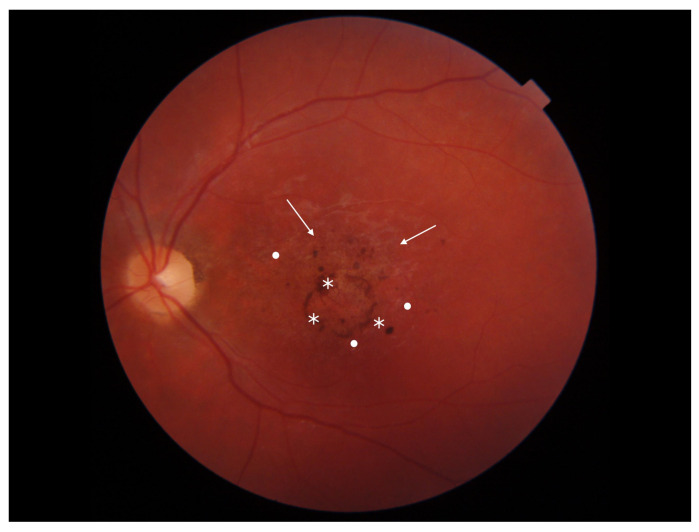
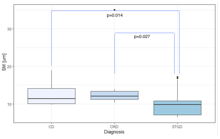
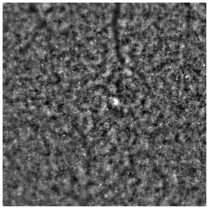
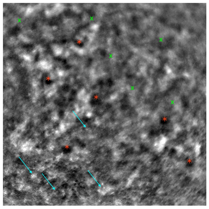
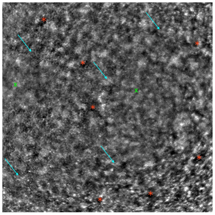
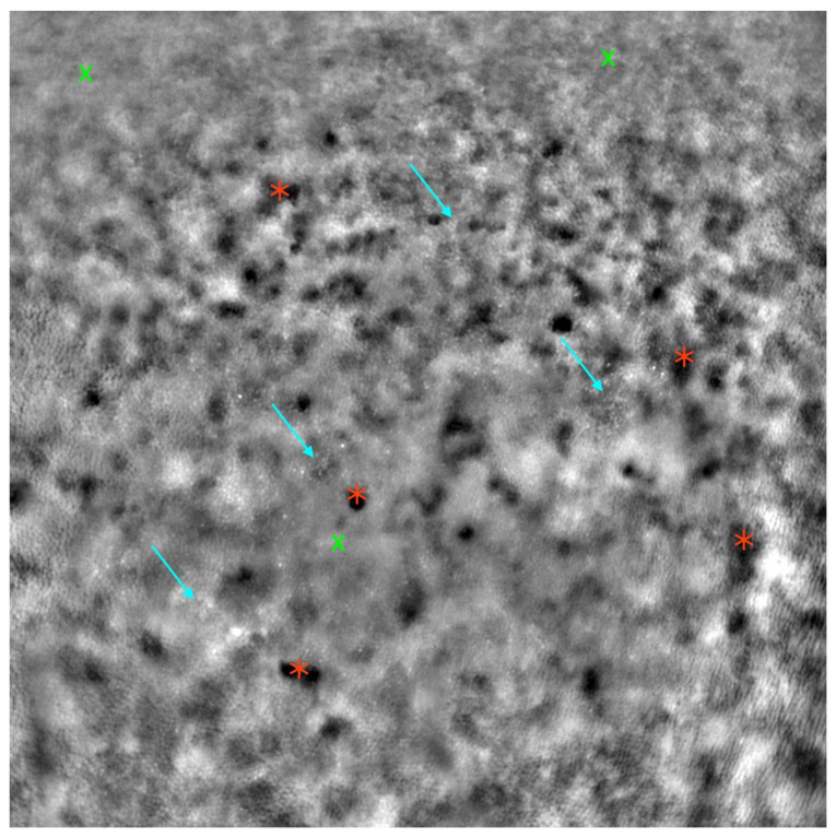
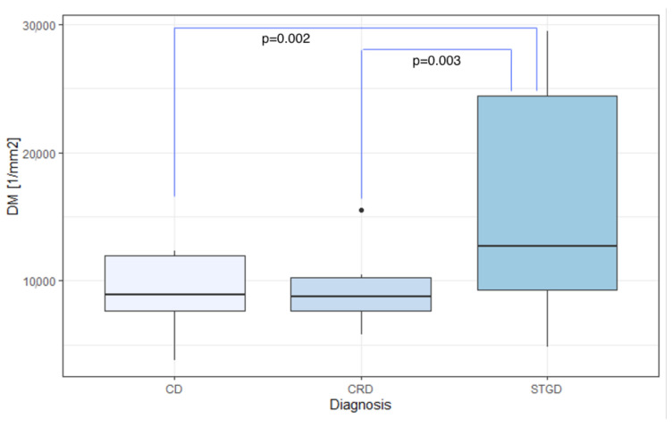
Inherited retinal dystrophies (IRDs) are genetic disorders that lead to the bilateral degeneration of the retina, causing irreversible vision loss. These conditions often manifest during the first and second decades of life, and their primary symptoms can be non-specific. Diagnostic processes encompass assessments of best-corrected visual acuity, fundoscopy, optical coherence tomography, fundus autofluorescence, fluorescein angiography, electrophysiological tests, and genetic testing. This study focuses on the application of adaptive optics (AO), a non-invasive retinal examination, for the assessment of patients with IRDs. AO facilitates the high-quality, detailed observation of retinal photoreceptor structures (cones and rods) and enables the quantitative analysis of parameters such as cone density (DM), cone spacing (SM), cone regularity (REG), and Voronoi analysis (N%6). AO examinations were conducted on eyes diagnosed with Stargardt disease (STGD, N=36), cone dystrophy (CD, N=9), and cone-rod dystrophy (CRD, N=8), and on healthy eyes (N=14). There were significant differences in the DM, SM, REG, and N%6 parameters between the healthy and IRD-affected eyes (p<0.001 for DM, SM, and REG; p=0.008 for N%6). The mean DM in the CD, CRD, and STGD groups was 8900.39/mm2, 9296.32/mm2, and 16,209.66/mm2, respectively, with a significant inter-group difference (p=0.006). The mean SM in the CD, CRD, and STGD groups was 12.37 μm, 14.82 μm, and 9.65 μm, respectively, with a significant difference observed between groups (p=0.002). However, no significant difference was found in REG and N%6 among the CD, CRD, and STGD groups. Significant differences were found in SM and DM between CD and STGD (p=0.014 for SM; p=0.003 for DM) and between CRD and STGD (p=0.027 for SM; p=0.003 for DM). Our findings suggest that AO holds significant potential as an impactful diagnostic tool for IRDs.

摘要

遗传性视网膜营养不良(IRDs)是一种遗传性疾病,可导致视网膜双侧变性,造成不可逆的视力丧失。这些病症通常在生命的第一个和第二个十年中出现,其主要症状可能不具有特异性。诊断过程包括最佳矫正视力评估、眼底镜检查、光学相干断层扫描、眼底自发荧光、荧光素血管造影、电生理测试和基因检测。本研究重点关注自适应光学(AO)这一非侵入性视网膜检查方法在IRD患者评估中的应用。AO有助于高质量、详细地观察视网膜光感受器结构(视锥细胞和视杆细胞),并能够对诸如视锥细胞密度(DM)、视锥细胞间距(SM)、视锥细胞规则性(REG)和Voronoi分析(N%6)等参数进行定量分析。对诊断为Stargardt病(STGD,N = 36)、视锥细胞营养不良(CD,N = 9)和视锥 - 视杆细胞营养不良(CRD,N = 8)的眼睛以及健康眼睛(N = 14)进行了AO检查。健康眼睛与受IRD影响的眼睛在DM、SM、REG和N%6参数上存在显著差异(DM、SM和REG的p<0.001;N%6的p = 0.008)。CD、CRD和STGD组的平均DM分别为8900.39/mm²、9296.32/mm²和16209.66/mm²,组间差异显著(p = 0.006)。CD、CRD和STGD组的平均SM分别为12.37μm、14.82μm和9.65μm,组间观察到显著差异(p = 0.002)。然而,CD、CRD和STGD组在REG和N%6方面未发现显著差异。CD与STGD之间以及CRD与STGD之间在SM和DM方面存在显著差异(SM的p = 0.014;DM的p = 0.003)。我们的研究结果表明,AO作为一种对IRD有重要影响的诊断工具具有巨大潜力。

https://cdn.ncbi.nlm.nih.gov/pmc/blobs/c787/10417470/01dc307623ba/diagnostics-13-02472-g009.jpg
https://cdn.ncbi.nlm.nih.gov/pmc/blobs/c787/10417470/c3feb70362a2/diagnostics-13-02472-g001.jpg
https://cdn.ncbi.nlm.nih.gov/pmc/blobs/c787/10417470/b591e2b244cc/diagnostics-13-02472-g002.jpg
https://cdn.ncbi.nlm.nih.gov/pmc/blobs/c787/10417470/8bd01c3ebec8/diagnostics-13-02472-g003.jpg
https://cdn.ncbi.nlm.nih.gov/pmc/blobs/c787/10417470/b167993d95ab/diagnostics-13-02472-g004.jpg
https://cdn.ncbi.nlm.nih.gov/pmc/blobs/c787/10417470/c0ddc40b3dec/diagnostics-13-02472-g005.jpg
https://cdn.ncbi.nlm.nih.gov/pmc/blobs/c787/10417470/bc597bc6afd2/diagnostics-13-02472-g006.jpg
https://cdn.ncbi.nlm.nih.gov/pmc/blobs/c787/10417470/7e7ae065e1c8/diagnostics-13-02472-g007.jpg
https://cdn.ncbi.nlm.nih.gov/pmc/blobs/c787/10417470/85e6c20162e9/diagnostics-13-02472-g008.jpg
https://cdn.ncbi.nlm.nih.gov/pmc/blobs/c787/10417470/01dc307623ba/diagnostics-13-02472-g009.jpg

相似文献

1
Characteristics of Rare Inherited Retinal Dystrophies in Adaptive Optics-A Study on 53 Eyes.自适应光学下罕见遗传性视网膜营养不良的特征——一项对53只眼的研究
Diagnostics (Basel). 2023 Jul 25;13(15):2472. doi: 10.3390/diagnostics13152472.
2
Progression of Rare Inherited Retinal Dystrophies May Be Monitored by Adaptive Optics Imaging.罕见遗传性视网膜营养不良的进展可通过自适应光学成像进行监测。
Life (Basel). 2023 Sep 5;13(9):1871. doi: 10.3390/life13091871.
3
Retinal Vascular Abnormalities in Different Types of Inherited Retinal Dystrophies Assessed by Optical Coherence Tomography Angiography.通过光学相干断层扫描血管造影评估不同类型遗传性视网膜营养不良中的视网膜血管异常。
J Curr Ophthalmol. 2021 Jul 5;33(2):189-196. doi: 10.4103/joco.joco_11_21. eCollection 2021 Apr-Jun.
4
High-resolution images in macular disorders.黄斑病变的高分辨率图像。
Rom J Ophthalmol. 2021 Apr-Jun;65(2):204-211. doi: 10.22336/rjo.2021.41.
5
Retinal Oxygenation in Inherited Diseases of the Retina.遗传性视网膜疾病中的视网膜氧合作用。
Genes (Basel). 2021 Feb 14;12(2):272. doi: 10.3390/genes12020272.
6
[Multimodal Approaches for the Analysis of Retinal Functional Disorders―Focusing on Retinal Detachment].[视网膜功能障碍分析的多模态方法——聚焦于视网膜脱离]
Nippon Ganka Gakkai Zasshi. 2017 Mar;121(3):185-231.
7
High-resolution imaging with adaptive optics in patients with inherited retinal degeneration.遗传性视网膜变性患者的自适应光学高分辨率成像。
Invest Ophthalmol Vis Sci. 2007 Jul;48(7):3283-91. doi: 10.1167/iovs.06-1422.
8
High-resolution retinal imaging of cone-rod dystrophy.视锥-视杆营养不良的高分辨率视网膜成像
Ophthalmology. 2006 Jun;113(6):1019.e1. doi: 10.1016/j.ophtha.2006.01.056. Epub 2006 May 2.
9
Phenotypic diversity in autosomal-dominant cone-rod dystrophy elucidated by adaptive optics retinal imaging.自适应光学视网膜成像揭示常染色体显性锥杆营养不良的表型多样性。
Br J Ophthalmol. 2018 Jan;102(1):136-141. doi: 10.1136/bjophthalmol-2017-310498. Epub 2017 Oct 26.
10
-Related Cone-Rod Dystrophy: Clinical Characteristics, Imaging Findings, and Genetic Test Results-A Case Report.相关 Cone-Rod 营养不良:临床特征、影像学发现和基因检测结果——病例报告。
Medicina (Kaunas). 2023 Feb 17;59(2):399. doi: 10.3390/medicina59020399.

引用本文的文献

1
Update on Clinical Trial Endpoints in Gene Therapy Trials for Inherited Retinal Diseases.遗传性视网膜疾病基因治疗试验的临床试验终点更新
J Clin Med. 2024 Sep 18;13(18):5512. doi: 10.3390/jcm13185512.
2
Modeling inherited retinal diseases using human induced pluripotent stem cell derived photoreceptor cells and retinal pigment epithelial cells.利用人类诱导多能干细胞衍生的光感受器细胞和视网膜色素上皮细胞对遗传性视网膜疾病进行建模。
Front Med (Lausanne). 2024 Jul 1;11:1328474. doi: 10.3389/fmed.2024.1328474. eCollection 2024.
3
Progression of Rare Inherited Retinal Dystrophies May Be Monitored by Adaptive Optics Imaging.

本文引用的文献

1
Evolution of adaptive optics retinal imaging [Invited].自适应光学视网膜成像的发展[特邀报告]
Biomed Opt Express. 2023 Feb 28;14(3):1307-1338. doi: 10.1364/BOE.485371. eCollection 2023 Mar 1.
2
Gene-agnostic therapeutic approaches for inherited retinal degenerations.针对遗传性视网膜变性的基因非特异性治疗方法。
Front Mol Neurosci. 2023 Jan 9;15:1068185. doi: 10.3389/fnmol.2022.1068185. eCollection 2022.
3
Twenty-five years of clinical applications using adaptive optics ophthalmoscopy [Invited].使用自适应光学检眼镜的25年临床应用[特邀报告]
罕见遗传性视网膜营养不良的进展可通过自适应光学成像进行监测。
Life (Basel). 2023 Sep 5;13(9):1871. doi: 10.3390/life13091871.
Biomed Opt Express. 2022 Dec 20;14(1):387-428. doi: 10.1364/BOE.472274. eCollection 2023 Jan 1.
4
RNA-based therapies in inherited retinal diseases.基于RNA的遗传性视网膜疾病治疗方法。
Ther Adv Ophthalmol. 2022 Nov 4;14:25158414221134602. doi: 10.1177/25158414221134602. eCollection 2022 Jan-Dec.
5
Newer therapeutic options for inherited retinal diseases: Gene and cell replacement therapy.遗传性视网膜疾病的新治疗选择:基因和细胞替代疗法。
Indian J Ophthalmol. 2022 Jul;70(7):2316-2325. doi: 10.4103/ijo.IJO_82_22.
6
Comparison of Cone Mosaic Metrics From Images Acquired With the SPECTRALIS High Magnification Module and Adaptive Optics Scanning Light Ophthalmoscopy.高倍模块光谱仪图像与自适应光学扫描检眼镜获取的锥细胞马赛克指标的比较。
Transl Vis Sci Technol. 2022 May 2;11(5):19. doi: 10.1167/tvst.11.5.19.
7
A Systematic Review of Artificial Intelligence Applications Used for Inherited Retinal Disease Management.人工智能在遗传性视网膜疾病管理中的应用:系统综述
Medicina (Kaunas). 2022 Mar 31;58(4):504. doi: 10.3390/medicina58040504.
8
The role of multimodal imaging and vision function testing in -related retinopathies and their relevance to future therapeutic interventions.多模态成像和视觉功能测试在相关视网膜病变中的作用及其与未来治疗干预的相关性。
Ther Adv Ophthalmol. 2021 Dec 19;13:25158414211056384. doi: 10.1177/25158414211056384. eCollection 2021 Jan-Dec.
9
Short-Term Parafoveal Cone Loss Despite Preserved Ellipsoid Zone in Rod Cone Dystrophy.尽管在视杆视锥营养不良中保留了椭圆体带,但短期旁中心锥细胞丢失。
Transl Vis Sci Technol. 2021 Dec 1;10(14):11. doi: 10.1167/tvst.10.14.11.
10
cGMP-grade human iPSC-derived retinal photoreceptor precursor cells rescue cone photoreceptor damage in non-human primates.cGMP 级人诱导多能干细胞衍生的视网膜光感受器前体细胞可挽救非人灵长类动物的圆锥体细胞光感受器损伤。
Stem Cell Res Ther. 2021 Aug 19;12(1):464. doi: 10.1186/s13287-021-02539-8.