• 文献检索
  • 文档翻译
  • 深度研究
  • 学术资讯
  • Suppr Zotero 插件Zotero 插件
  • 邀请有礼
  • 套餐&价格
  • 历史记录
应用&插件
Suppr Zotero 插件Zotero 插件浏览器插件Mac 客户端Windows 客户端微信小程序
定价
高级版会员购买积分包购买API积分包
服务
文献检索文档翻译深度研究API 文档MCP 服务
关于我们
关于 Suppr公司介绍联系我们用户协议隐私条款
关注我们

Suppr 超能文献

核心技术专利:CN118964589B侵权必究
粤ICP备2023148730 号-1Suppr @ 2026

文献检索

告别复杂PubMed语法,用中文像聊天一样搜索,搜遍4000万医学文献。AI智能推荐,让科研检索更轻松。

立即免费搜索

文件翻译

保留排版,准确专业,支持PDF/Word/PPT等文件格式,支持 12+语言互译。

免费翻译文档

深度研究

AI帮你快速写综述,25分钟生成高质量综述,智能提取关键信息,辅助科研写作。

立即免费体验

自适应光学视网膜成像揭示常染色体显性锥杆营养不良的表型多样性。

Phenotypic diversity in autosomal-dominant cone-rod dystrophy elucidated by adaptive optics retinal imaging.

作者信息

Song Hongxin, Rossi Ethan A, Stone Edwin, Latchney Lisa, Williams David, Dubra Alfredo, Chung Mina

机构信息

Beijing Institute of Ophthalmology, Beijing Tongren Eye Center, Beijing Tongren Hospital, Capital Medical University, Beijing Key Laboratory of Ophthalmology and Visual SciencesNational Engineering Research Center for Ophthalmic Equipment, Beijing, China.

University of Rochester, Center for Visual Science, Rochester, New York, USA.

出版信息

Br J Ophthalmol. 2018 Jan;102(1):136-141. doi: 10.1136/bjophthalmol-2017-310498. Epub 2017 Oct 26.

DOI:10.1136/bjophthalmol-2017-310498
PMID:29074494
原文链接:https://pmc.ncbi.nlm.nih.gov/articles/PMC5754866/
Abstract

PURPOSE

Several genes causing autosomal-dominant cone-rod dystrophy (AD-CRD) have been identified. However, the mechanisms by which genetic mutations lead to cellular loss in human disease remain poorly understood. Here we combine genotyping with high-resolution adaptive optics retinal imaging to elucidate the retinal phenotype at a cellular level in patients with AD-CRD harbouring a defect in the gene.

METHODS

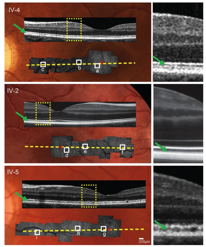
Nine affected members of a four-generation AD-CRD pedigree and three unaffected first-degree relatives underwent clinical examinations including visual acuity, fundus examination, Goldmann perimetry, spectral domain optical coherence tomography and electroretinography. Genome-wide scan followed by bidirectional sequencing was performed on all affected participants. High-resolution imaging using a custom adaptive optics scanning light ophthalmoscope (AOSLO) was performed for selected participants.

RESULTS

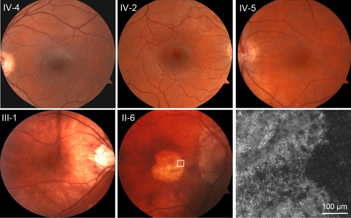
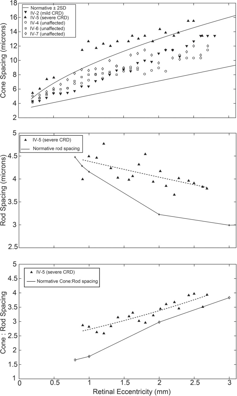
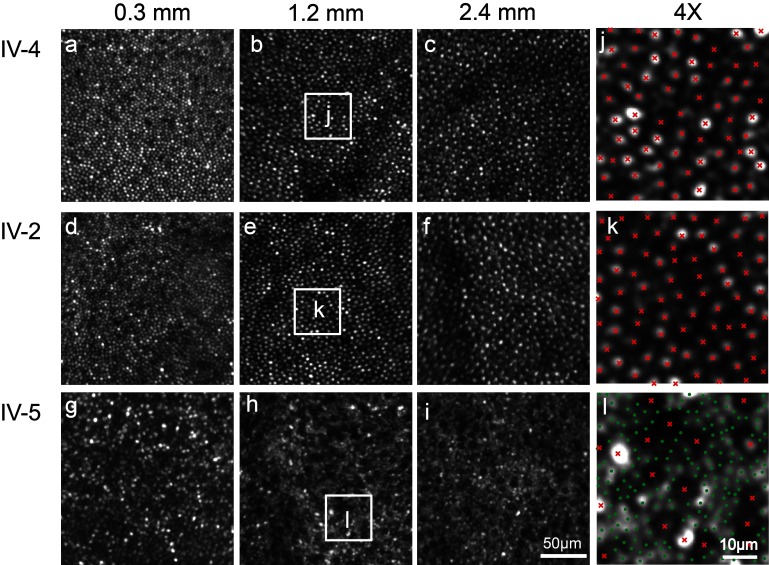
Clinical evaluations showed a range of disease severity from normal fundus appearance in teenaged patients to pronounced macular atrophy in older patients. Molecular genetic testing showed a mutation in in segregating with disease. AOSLO imaging revealed that of the two teenage patients with mild disease, one had severe disruption of the photoreceptor mosaic while the other had a normal cone mosaic.

CONCLUSIONS

AOSLO imaging demonstrated variability in the pattern of cone and rod cell loss between two teenage cousins with early AD-CRD, who had similar clinical features and had the identical disease-causing mutation in . This finding suggests that a mutation in does not lead to the same degree of AD-CRD in all patients. Modifying factors may mitigate or augment disease severity, leading to different retinal cellular phenotypes.

摘要

目的

已鉴定出几种导致常染色体显性遗传性锥杆营养不良(AD-CRD)的基因。然而,基因突变导致人类疾病中细胞丢失的机制仍知之甚少。在此,我们将基因分型与高分辨率自适应光学视网膜成像相结合,以在细胞水平上阐明携带该基因缺陷的AD-CRD患者的视网膜表型。

方法

对一个四代AD-CRD家系的9名患病成员和3名未患病的一级亲属进行了临床检查,包括视力、眼底检查、Goldmann视野检查、光谱域光学相干断层扫描和视网膜电图检查。对所有患病参与者进行全基因组扫描,随后进行双向测序。对选定的参与者使用定制的自适应光学扫描激光检眼镜(AOSLO)进行高分辨率成像。

结果

临床评估显示疾病严重程度范围较广,从青少年患者眼底外观正常到老年患者明显的黄斑萎缩。分子遗传学检测显示该基因存在与疾病共分离的突变。AOSLO成像显示,在两名患有轻度疾病的青少年患者中,一名患者的光感受器镶嵌严重破坏,而另一名患者的视锥镶嵌正常。

结论

AOSLO成像显示,两名患有早期AD-CRD的青少年表亲,尽管临床特征相似且致病突变相同,但视锥和视杆细胞丢失模式存在差异。这一发现表明,该基因突变在所有患者中不会导致相同程度的AD-CRD。修饰因子可能会减轻或加重疾病严重程度,导致不同的视网膜细胞表型。

https://cdn.ncbi.nlm.nih.gov/pmc/blobs/1757/5754866/1e9f7f085d31/bjophthalmol-2017-310498f04.jpg
https://cdn.ncbi.nlm.nih.gov/pmc/blobs/1757/5754866/7590a3921219/bjophthalmol-2017-310498f01.jpg
https://cdn.ncbi.nlm.nih.gov/pmc/blobs/1757/5754866/8f2476d18c9d/bjophthalmol-2017-310498f02.jpg
https://cdn.ncbi.nlm.nih.gov/pmc/blobs/1757/5754866/095ef9e81ae6/bjophthalmol-2017-310498f03.jpg
https://cdn.ncbi.nlm.nih.gov/pmc/blobs/1757/5754866/1e9f7f085d31/bjophthalmol-2017-310498f04.jpg

相似文献

1
Phenotypic diversity in autosomal-dominant cone-rod dystrophy elucidated by adaptive optics retinal imaging.自适应光学视网膜成像揭示常染色体显性锥杆营养不良的表型多样性。
Br J Ophthalmol. 2018 Jan;102(1):136-141. doi: 10.1136/bjophthalmol-2017-310498. Epub 2017 Oct 26.
2
Cone dystrophy or macular dystrophy associated with novel autosomal dominant mutations.与新型常染色体显性突变相关的视锥营养不良或黄斑营养不良。
Mol Vis. 2017 Apr 3;23:198-209. eCollection 2017.
3
High-resolution retinal imaging of cone-rod dystrophy.视锥-视杆营养不良的高分辨率视网膜成像
Ophthalmology. 2006 Jun;113(6):1019.e1. doi: 10.1016/j.ophtha.2006.01.056. Epub 2006 May 2.
4
Characterization of the cone-rod dystrophy retinal phenotype caused by novel homozygous DRAM2 mutations.新型 DRAM2 基因突变致锥杆细胞营养不良的视网膜表型特征。
Exp Eye Res. 2019 Oct;187:107752. doi: 10.1016/j.exer.2019.107752. Epub 2019 Aug 5.
5
GUCY2D- or GUCA1A-related autosomal dominant cone-rod dystrophy: is there a phenotypic difference?与GUCY2D或GUCA1A相关的常染色体显性遗传性视锥视杆营养不良:是否存在表型差异?
Retina. 2014 Aug;34(8):1576-87. doi: 10.1097/IAE.0000000000000129.
6
High-resolution adaptive optics retinal imaging of cellular structure in choroideremia.无脉络膜症中细胞结构的高分辨率自适应光学视网膜成像。
Invest Ophthalmol Vis Sci. 2014 Sep 4;55(10):6381-97. doi: 10.1167/iovs.13-13454.
7
A ROD-CONE DYSTROPHY IS SYSTEMATICALLY ASSOCIATED TO THE RTN4IP1 RECESSIVE OPTIC ATROPHY.一种 Rod-Cone 营养不良与 RTN4IP1 隐性视神经萎缩有系统关联。
Retina. 2021 Aug 1;41(8):1771-1779. doi: 10.1097/IAE.0000000000003054.
8
Characterization of GUCA1A-associated dominant cone/cone-rod dystrophy: low prevalence among Japanese patients with inherited retinal dystrophies.GUCA1A 相关性显性视锥/视锥-杆营养不良的特征:日本遗传性视网膜病变患者中的低患病率。
Sci Rep. 2019 Nov 14;9(1):16851. doi: 10.1038/s41598-019-52660-1.
9
Maternal uniparental isodisomy of chromosome 6 reveals a TULP1 mutation as a novel cause of cone dysfunction.母源单亲二体性 6 号染色体导致视锥细胞功能障碍,揭示 TULP1 突变是其新的致病原因。
Ophthalmology. 2013 Jun;120(6):1239-46. doi: 10.1016/j.ophtha.2012.12.005. Epub 2013 Mar 15.
10
Ophthalmic features of cone-rod dystrophy caused by pathogenic variants in the ALMS1 gene.ALMS1 基因突变致锥-杆细胞营养不良的眼部特征。
Acta Ophthalmol. 2018 Jun;96(4):e445-e454. doi: 10.1111/aos.13612. Epub 2017 Nov 30.

引用本文的文献

1
Molecular Mechanisms Governing Sight Loss in Inherited Cone Disorders.遗传性锥细胞疾病致盲的分子机制。
Genes (Basel). 2024 Jun 1;15(6):727. doi: 10.3390/genes15060727.
2
LONGITUDINAL ADAPTIVE OPTICS SCANNING LASER OPHTHALMOSCOPY REVEALS REGIONAL VARIATION IN CONE AND ROD PHOTORECEPTOR LOSS IN STARGARDT DISEASE.纵向自适应光学扫描激光检眼镜显示斯塔加特病中视锥和视杆光感受器的丢失存在区域性差异。
Retina. 2024 Aug 1;44(8):1403-1412. doi: 10.1097/IAE.0000000000004104.
3
Characteristics of Rare Inherited Retinal Dystrophies in Adaptive Optics-A Study on 53 Eyes.

本文引用的文献

1
The Negative Cone Mosaic: A New Manifestation of the Optical Stiles-Crawford Effect in Normal Eyes.负锥镶嵌:正常眼睛中光学斯泰尔斯-克劳福德效应的一种新表现形式。
Invest Ophthalmol Vis Sci. 2015 Nov;56(12):7043-50. doi: 10.1167/iovs.15-17022.
2
Cone and rod loss in Stargardt disease revealed by adaptive optics scanning light ophthalmoscopy.自适应光学扫描激光检眼镜揭示的斯塔加特病中的视锥和视杆细胞丢失
JAMA Ophthalmol. 2015 Oct;133(10):1198-203. doi: 10.1001/jamaophthalmol.2015.2443.
3
Fluorescence adaptive optics scanning laser ophthalmoscope for detection of reduced cones and hypoautofluorescent spots in fundus albipunctatus.
自适应光学下罕见遗传性视网膜营养不良的特征——一项对53只眼的研究
Diagnostics (Basel). 2023 Jul 25;13(15):2472. doi: 10.3390/diagnostics13152472.
4
Pearls and Pitfalls of Adaptive Optics Ophthalmoscopy in Inherited Retinal Diseases.遗传性视网膜疾病中自适应光学检眼镜检查的要点与陷阱
Diagnostics (Basel). 2023 Jul 19;13(14):2413. doi: 10.3390/diagnostics13142413.
5
Extracting spacing-derived estimates of rod density in healthy retinae.提取健康视网膜中基于间距的视杆细胞密度估计值。
Biomed Opt Express. 2022 Dec 5;14(1):1-17. doi: 10.1364/BOE.473101. eCollection 2023 Jan 1.
6
Twenty-five years of clinical applications using adaptive optics ophthalmoscopy [Invited].使用自适应光学检眼镜的25年临床应用[特邀报告]
Biomed Opt Express. 2022 Dec 20;14(1):387-428. doi: 10.1364/BOE.472274. eCollection 2023 Jan 1.
7
Evaluation of retinal function and morphology in isoametropic amblyopia.评价屈光不正性弱视的视网膜功能和形态。
Indian J Ophthalmol. 2023 Jan;71(1):222-228. doi: 10.4103/ijo.IJO_1685_22.
8
Short-term Assessment of Subfoveal Injection of Adeno-Associated Virus-Mediated hCHM Gene Augmentation in Choroideremia Using Adaptive Optics Ophthalmoscopy.应用自适应光学眼底镜评估腺相关病毒介导的 hCHM 基因增强在脉络膜黑蒙患者眼内脉络膜注射的短期疗效。
JAMA Ophthalmol. 2022 Apr 1;140(4):411-420. doi: 10.1001/jamaophthalmol.2022.0158.
9
Patches of Dysflective Cones in Eyes With No Known Disease.眼部无已知疾病的失谐锥体斑。
Invest Ophthalmol Vis Sci. 2022 Jan 3;63(1):29. doi: 10.1167/iovs.63.1.29.
10
Retinal imaging in inherited retinal diseases.遗传性视网膜疾病中的视网膜成像。
Ann Eye Sci. 2020 Sep;5. doi: 10.21037/aes-20-81. Epub 2020 Sep 15.
用于检测白点状眼底病变中视锥细胞减少和低自发荧光斑的荧光自适应光学扫描激光检眼镜
JAMA Ophthalmol. 2014 Sep;132(9):1099-104. doi: 10.1001/jamaophthalmol.2014.1079.
4
In vivo imaging of human cone photoreceptor inner segments.人眼视锥细胞内节的活体成像。
Invest Ophthalmol Vis Sci. 2014 Jun 6;55(7):4244-51. doi: 10.1167/iovs.14-14542.
5
Observation of cone and rod photoreceptors in normal subjects and patients using a new generation adaptive optics scanning laser ophthalmoscope.使用新一代自适应光学扫描激光检眼镜对正常受试者和患者的视锥和视杆光感受器进行观察。
Biomed Opt Express. 2011 Aug 1;2(8):2189-201. doi: 10.1364/BOE.2.002189. Epub 2011 Jul 8.
6
Photoreceptor structure and function in patients with congenital achromatopsia.先天性色觉缺失患者的感光器结构和功能。
Invest Ophthalmol Vis Sci. 2011 Sep 21;52(10):7298-308. doi: 10.1167/iovs.11-7762.
7
Noninvasive imaging of the human rod photoreceptor mosaic using a confocal adaptive optics scanning ophthalmoscope.使用共焦自适应光学扫描检眼镜对人类视杆光感受器镶嵌进行无创成像。
Biomed Opt Express. 2011 Jul 1;2(7):1864-76. doi: 10.1364/BOE.2.001864. Epub 2011 Jun 8.
8
Variation of cone photoreceptor packing density with retinal eccentricity and age.锥形光感受器密度随视网膜偏心度和年龄的变化。
Invest Ophthalmol Vis Sci. 2011 Sep 21;52(10):7376-84. doi: 10.1167/iovs.11-7199. Print 2011 Sep.
9
In vivo imaging of the human rod photoreceptor mosaic.人眼视杆细胞外节的活体成像。
Opt Lett. 2011 Jan 1;36(1):31-3. doi: 10.1364/OL.36.000031.
10
Longitudinal study of cone photoreceptors during retinal degeneration and in response to ciliary neurotrophic factor treatment.视网膜变性过程中和睫状神经营养因子治疗反应中视锥细胞的纵向研究。
Invest Ophthalmol Vis Sci. 2011 Apr 6;52(5):2219-26. doi: 10.1167/iovs.10-6479.