Nephrology Unit- Kidney Histomorphology and Molecular Biology Laboratory, Department of Medicine-DIMED, University of Padua, Via Giustiniani, 2, 235128, Padua, Italy.
Nephrology Service, Hospital Británico de Buenos Aires, Buenos Aires, Argentina.
J Nephrol. 2023 Dec;36(9):2499-2506. doi: 10.1007/s40620-023-01725-6. Epub 2023 Aug 18.
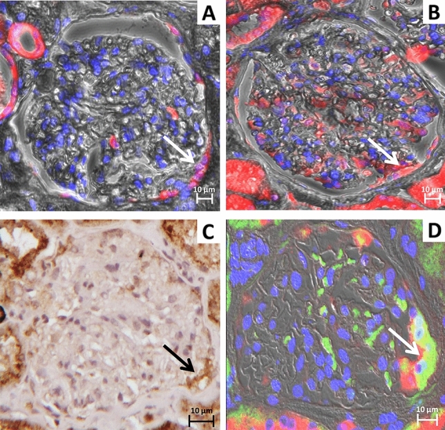
Parietal epithelial cells are a heterogeneous population of cells located on Bowman's capsule. These cells are known to internalize albumin with a still undetermined mechanism, although albumin has been shown to induce phenotypic changes in parietal epithelial cells. Proximal tubular cells are the main actors in albumin handling via the macromolecular complex composed by ClC-5, megalin, and cubilin. This study investigated the role of ClC-5, megalin, and cubilin in the parietal epithelial cells of kidney biopsies from proteinuric lupus nephritis patients and control subjects and identified phenotypical changes occurring in the pathological milieu.
Immunohistochemistry and immunofluorescence analyses for ClC-5, megalin, cubilin, ANXA3, podocalyxin, CD24, CD44, HSA, and LTA marker were performed on 23 kidney biopsies from patients with Lupus Nephritis and 9 control biopsies (obtained from nephrectomies for renal cancer).
Two sub-populations of hypertrophic parietal epithelial cells ANXA3/Podocalyxin/CD44, both expressing ClC-5, megalin, and cubilin and located at the tubular pole, were identified and characterized: the first one, CD24/HSA/LTA had characteristics of human adult parietal epithelial multipotent progenitors, the second one, CD24/LTA/HSA committed to become phenotypically proximal tubular cells. The number of glomeruli presenting hypertrophic parietal epithelial cells positive for ClC-5, megalin, and cubilin were significantly higher in lupus nephritis patients than in controls.
Our results may provide further insight into the role of hypertrophic parietal epithelial cells located at the tubular pole and their possible involvement in protein endocytosis in lupus nephritis patients. These data also suggest that the presence of hypertrophic parietal epithelial cells in Bowman's capsule represents a potential resource for responding to protein overload observed in other glomerulonephritis.
壁细胞是位于鲍曼囊的异质性细胞群体。尽管白蛋白已被证明会诱导壁细胞发生表型变化,但这些细胞内化白蛋白的机制仍未确定。近端肾小管细胞是通过由 ClC-5、巨球蛋白和 cubilin 组成的大分子复合物处理白蛋白的主要作用者。本研究调查了 ClC-5、巨球蛋白和 cubilin 在蛋白尿性狼疮肾炎患者和对照者的肾活检组织中的壁细胞中的作用,并确定了在病理环境中发生的表型变化。
对 23 例狼疮肾炎患者和 9 例对照患者(因肾癌行肾切除术获得)的肾活检组织进行 ClC-5、巨球蛋白、cubilin、ANXA3、足细胞、CD24、CD44、HSA 和 LTA 标志物的免疫组织化学和免疫荧光分析。
鉴定并表征了位于管状极的两种肥大壁细胞 ANXA3/Podocalyxin/CD44 的亚群:第一个亚群 CD24/HSA/LTA 具有人类成年壁细胞多能祖细胞的特征,第二个亚群 CD24/LTA/HSA 向表型近端肾小管细胞分化。ClC-5、巨球蛋白和 cubilin 阳性的肥大壁细胞的肾小球数量在狼疮肾炎患者中明显高于对照组。
我们的结果可能为位于管状极的肥大壁细胞的作用及其在狼疮肾炎患者中可能参与蛋白内吞作用提供进一步的认识。这些数据还表明,鲍曼囊中肥大壁细胞的存在代表了对其他肾小球肾炎中观察到的蛋白过载的一种潜在反应资源。