From the Russell H. Morgan Department of Radiology and Radiological Science (S.D., J.G.K., E.G., H.A.I., K.M., K.T., E.K.F.) and Department of Biomedical Engineering (W.B.Z.), Johns Hopkins University School of Medicine, 601 N Carolina St, Baltimore, MD 21287; Division of Musculoskeletal Imaging, Department of Radiology, Mayo Clinic, Rochester, Minn (F.I.B.); Department of Radiology, New York University Grossman School of Medicine, New York, NY (J.F.); Department of Radiology and Imaging, Hospital for Special Surgery, New York, NY (J.A.C.); and Department of Radiology, Quantitative Imaging Center, Boston University School of Medicine, Boston, Mass (A.G.).
Radiology. 2023 Aug;308(2):e230344. doi: 10.1148/radiol.230344.
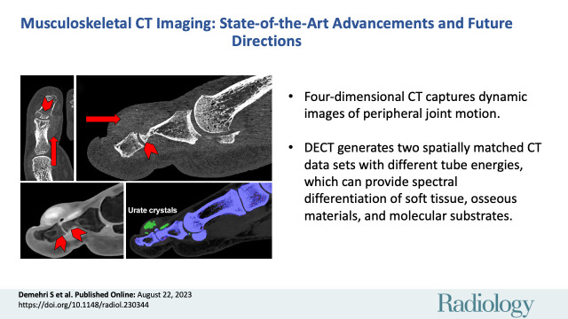
CT is one of the most widely used modalities for musculoskeletal imaging. Recent advancements in the field include the introduction of four-dimensional CT, which captures a CT image during motion; cone-beam CT, which uses flat-panel detectors to capture the lower extremities in weight-bearing mode; and dual-energy CT, which operates at two different x-ray potentials to improve the contrast resolution to facilitate the assessment of tissue material compositions such as tophaceous gout deposits and bone marrow edema. Most recently, photon-counting CT (PCCT) has been introduced. PCCT is a technique that uses photon-counting detectors to produce an image with higher spatial and contrast resolution than conventional multidetector CT systems. In addition, postprocessing techniques such as three-dimensional printing and cinematic rendering have used CT data to improve the generation of both physical and digital anatomic models. Last, advancements in the application of artificial intelligence to CT imaging have enabled the automatic evaluation of musculoskeletal pathologies. In this review, the authors discuss the current state of the above CT technologies, their respective advantages and disadvantages, and their projected future directions for various musculoskeletal applications.
CT 是肌肉骨骼成像中应用最广泛的模态之一。该领域的最新进展包括引入了四维 CT,它可以在运动过程中捕获 CT 图像;锥形束 CT,它使用平板探测器以承重模式捕获下肢;以及双能 CT,它在两个不同的 X 射线位操作以提高对比度分辨率,从而有助于评估组织材料成分,如痛风石沉积和骨髓水肿。最近,还引入了光子计数 CT(PCCT)。PCCT 是一种使用光子计数探测器生成图像的技术,与传统的多探测器 CT 系统相比,它具有更高的空间和对比度分辨率。此外,三维打印和电影渲染等后处理技术已经使用 CT 数据来改进物理和数字解剖模型的生成。最后,人工智能在 CT 成像中的应用的进步使得自动评估肌肉骨骼病变成为可能。在这篇综述中,作者讨论了上述 CT 技术的现状、各自的优缺点,以及它们在各种肌肉骨骼应用中的未来发展方向。