• 文献检索
  • 文档翻译
  • 深度研究
  • 学术资讯
  • Suppr Zotero 插件Zotero 插件
  • 邀请有礼
  • 套餐&价格
  • 历史记录
应用&插件
Suppr Zotero 插件Zotero 插件浏览器插件Mac 客户端Windows 客户端微信小程序
定价
高级版会员购买积分包购买API积分包
服务
文献检索文档翻译深度研究API 文档MCP 服务
关于我们
关于 Suppr公司介绍联系我们用户协议隐私条款
关注我们

Suppr 超能文献

核心技术专利:CN118964589B侵权必究
粤ICP备2023148730 号-1Suppr @ 2026

文献检索

告别复杂PubMed语法,用中文像聊天一样搜索,搜遍4000万医学文献。AI智能推荐,让科研检索更轻松。

立即免费搜索

文件翻译

保留排版,准确专业,支持PDF/Word/PPT等文件格式,支持 12+语言互译。

免费翻译文档

深度研究

AI帮你快速写综述,25分钟生成高质量综述,智能提取关键信息,辅助科研写作。

立即免费体验

人源 OPA1 调控线粒体膜重塑的结构机制

Structural mechanism of mitochondrial membrane remodelling by human OPA1.

机构信息

Medical Biochemistry & Molecular Biology, Center for Molecular Signaling, PZMS, Saarland University Medical School, Homburg, Germany.

Department of Biochemistry, University of Colorado Boulder, Boulder, CO, USA.

出版信息

Nature. 2023 Aug;620(7976):1101-1108. doi: 10.1038/s41586-023-06441-6. Epub 2023 Aug 23.

DOI:10.1038/s41586-023-06441-6
PMID:37612504
原文链接:https://pmc.ncbi.nlm.nih.gov/articles/PMC10875962/
Abstract

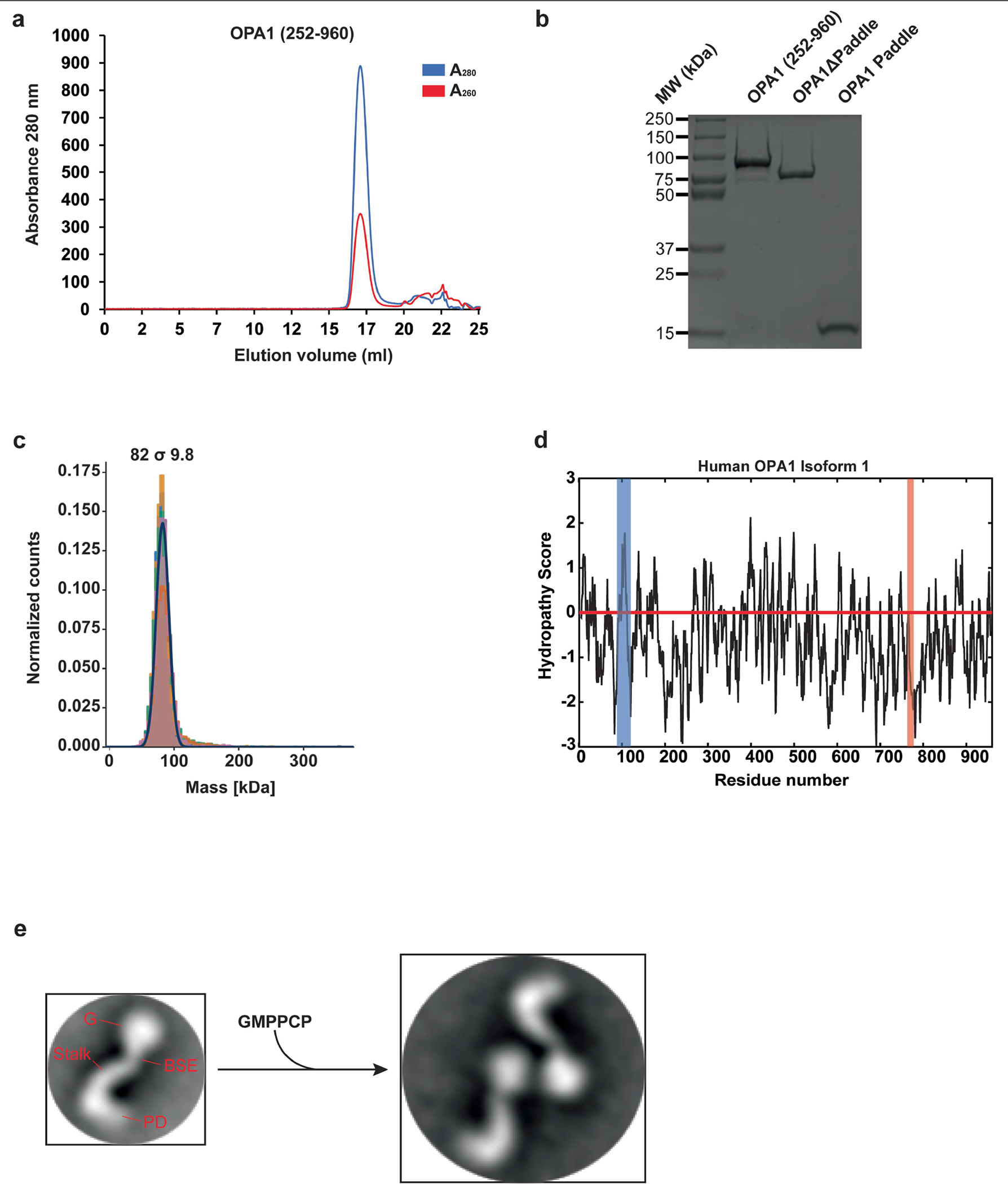
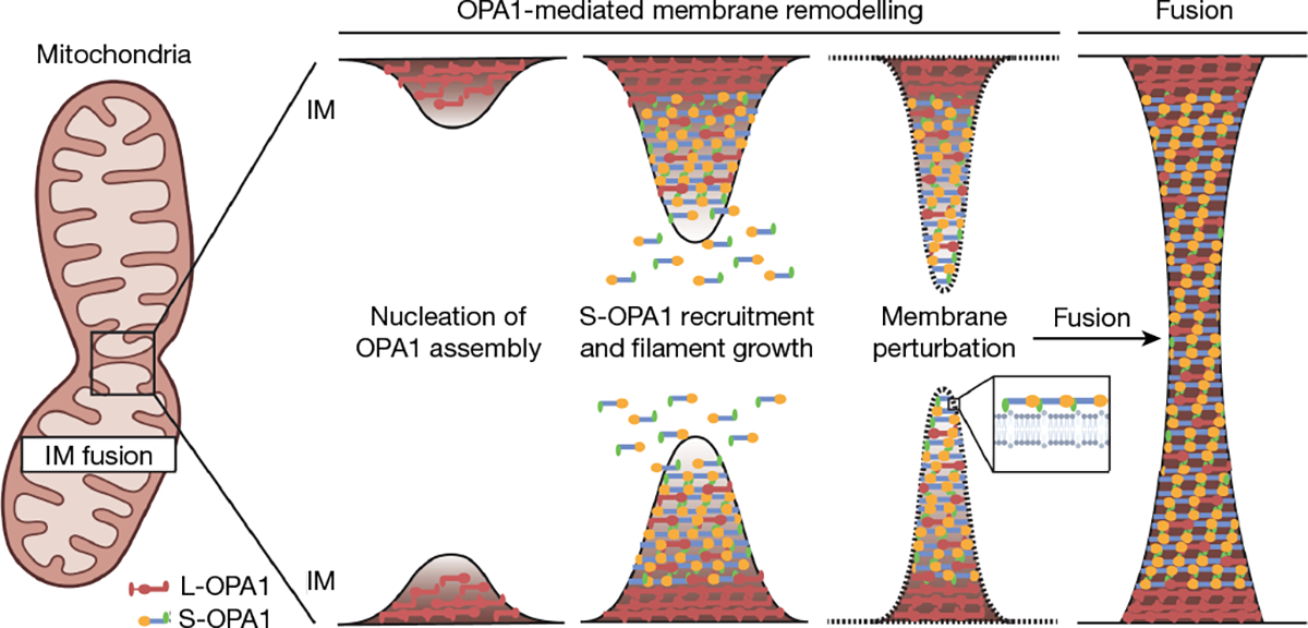
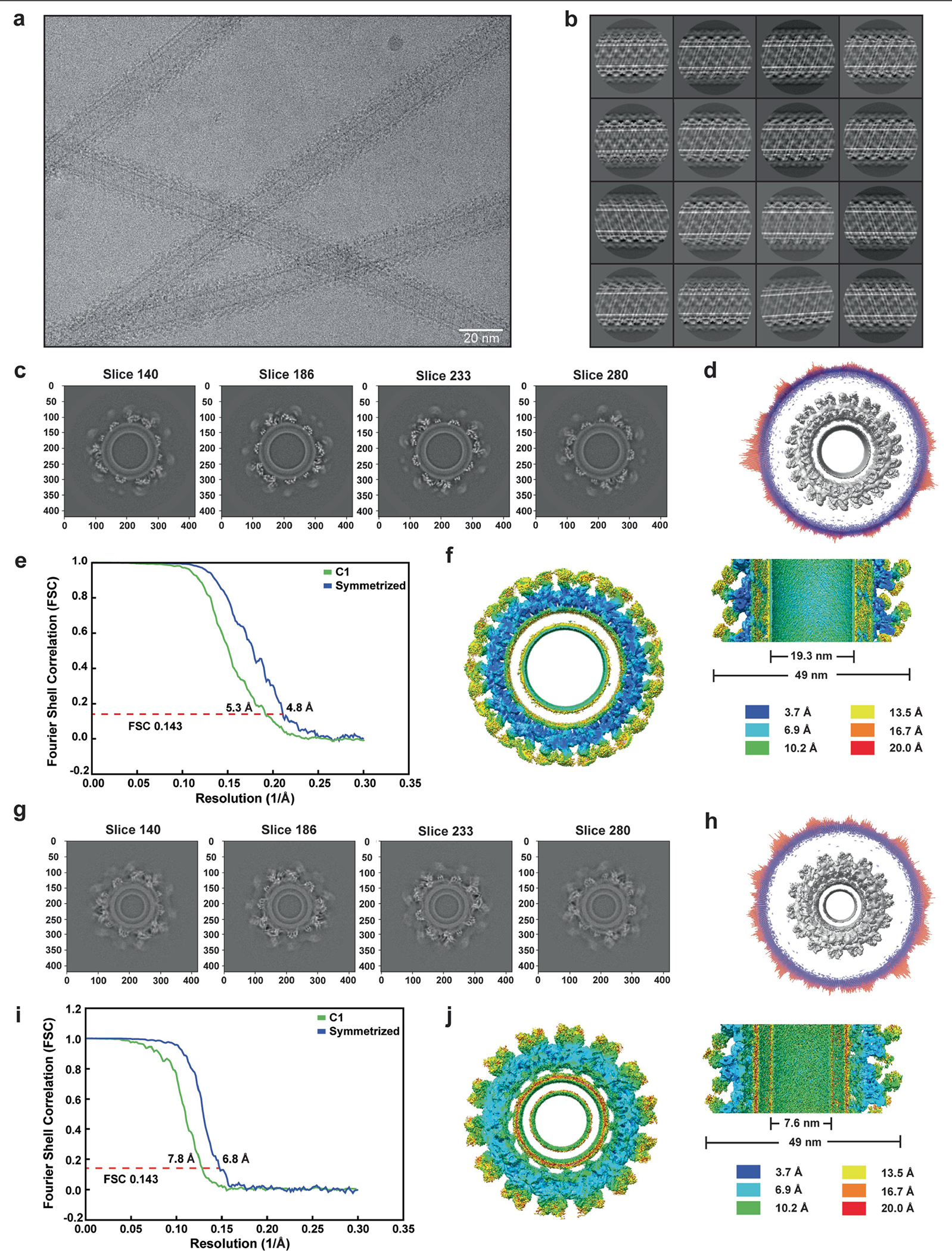
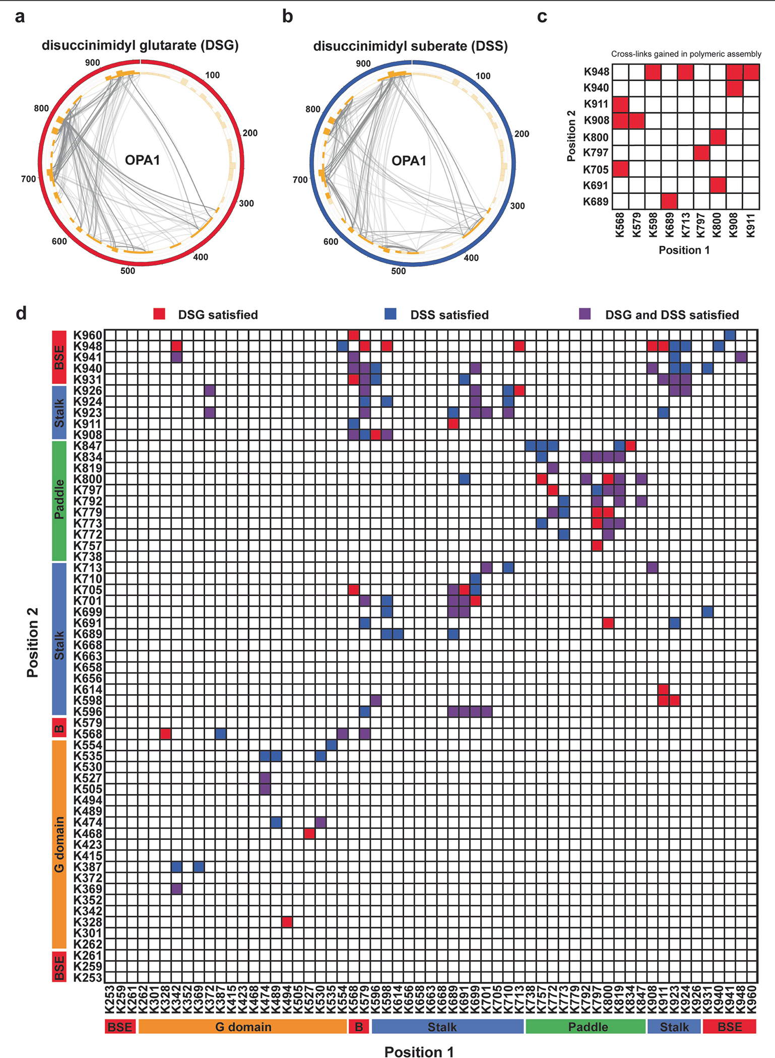
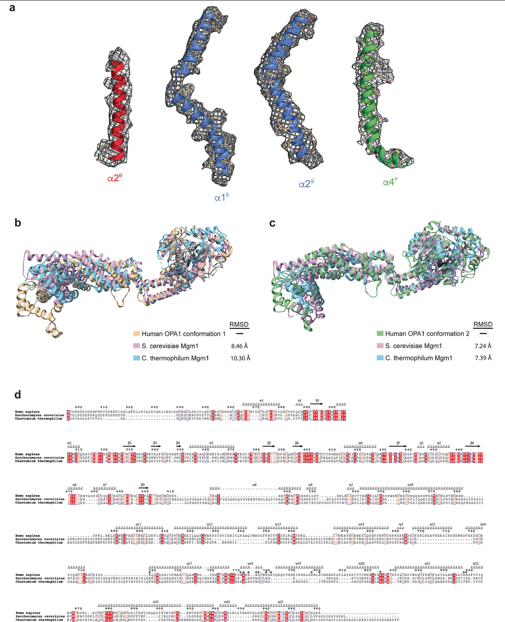
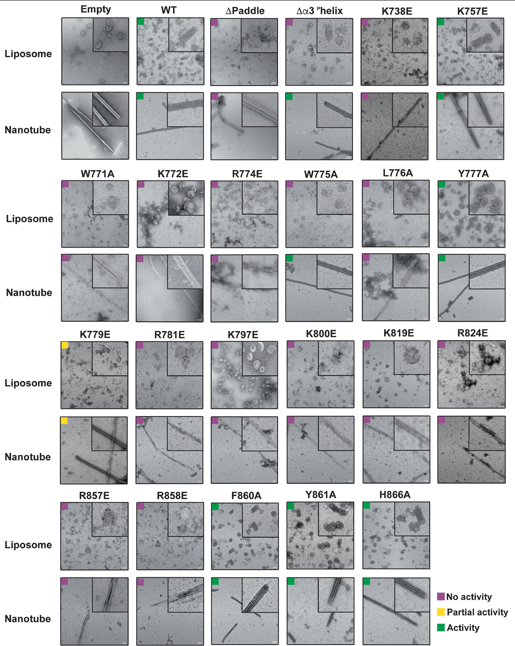
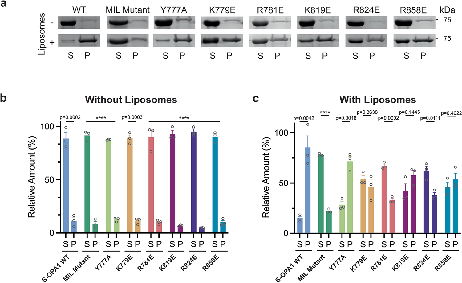
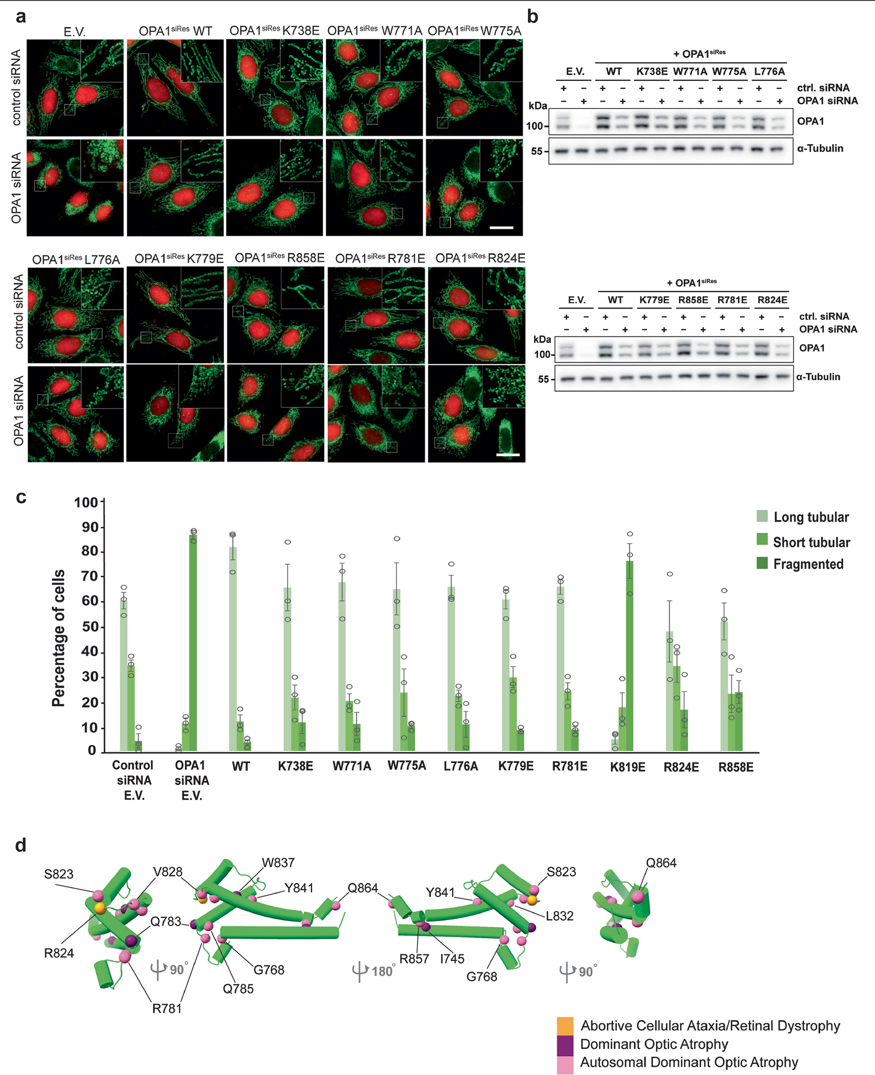
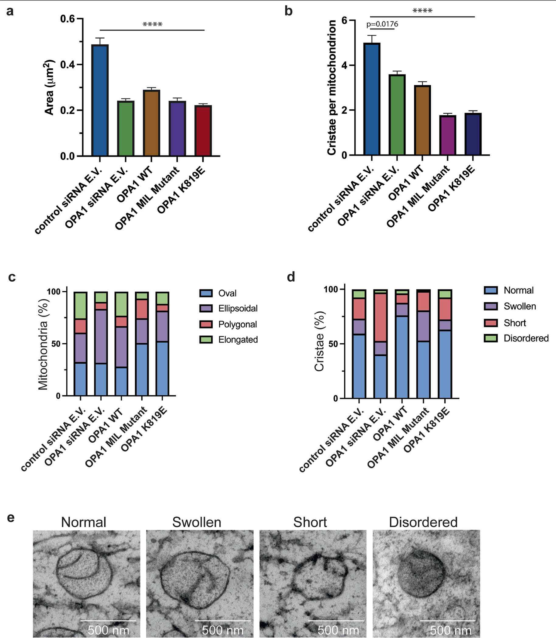
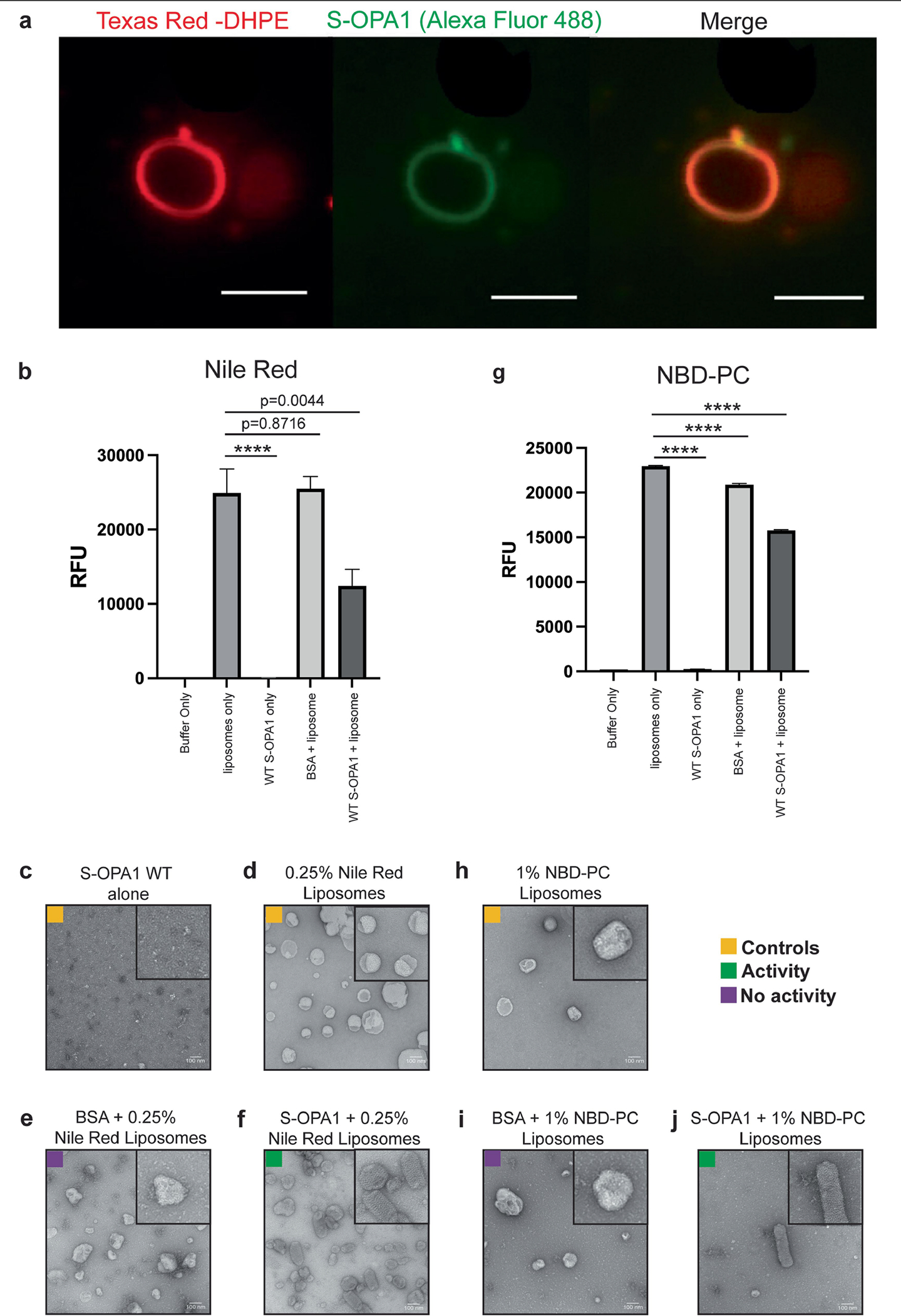
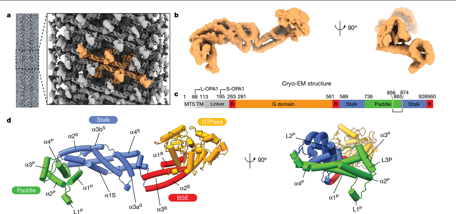
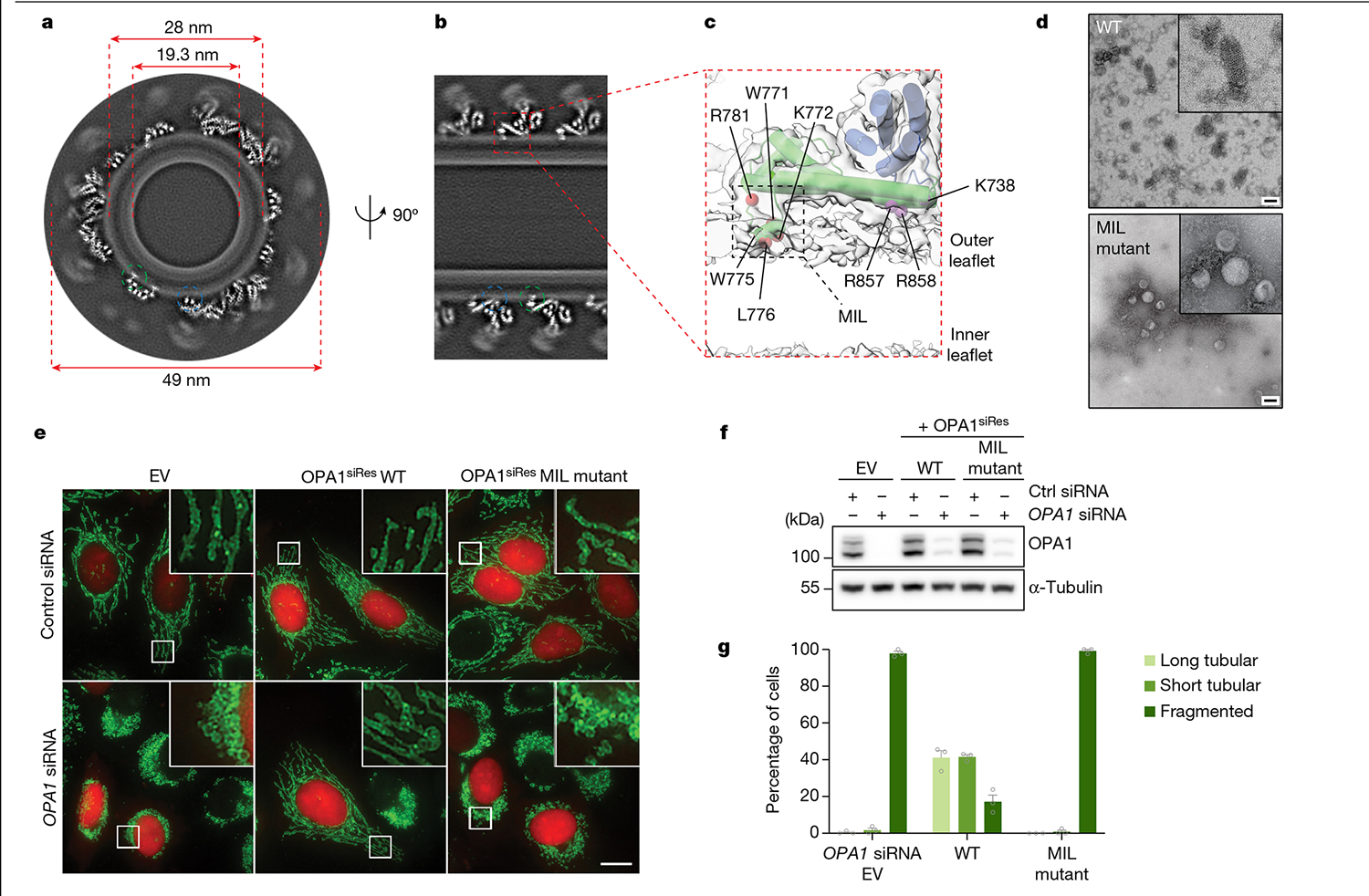
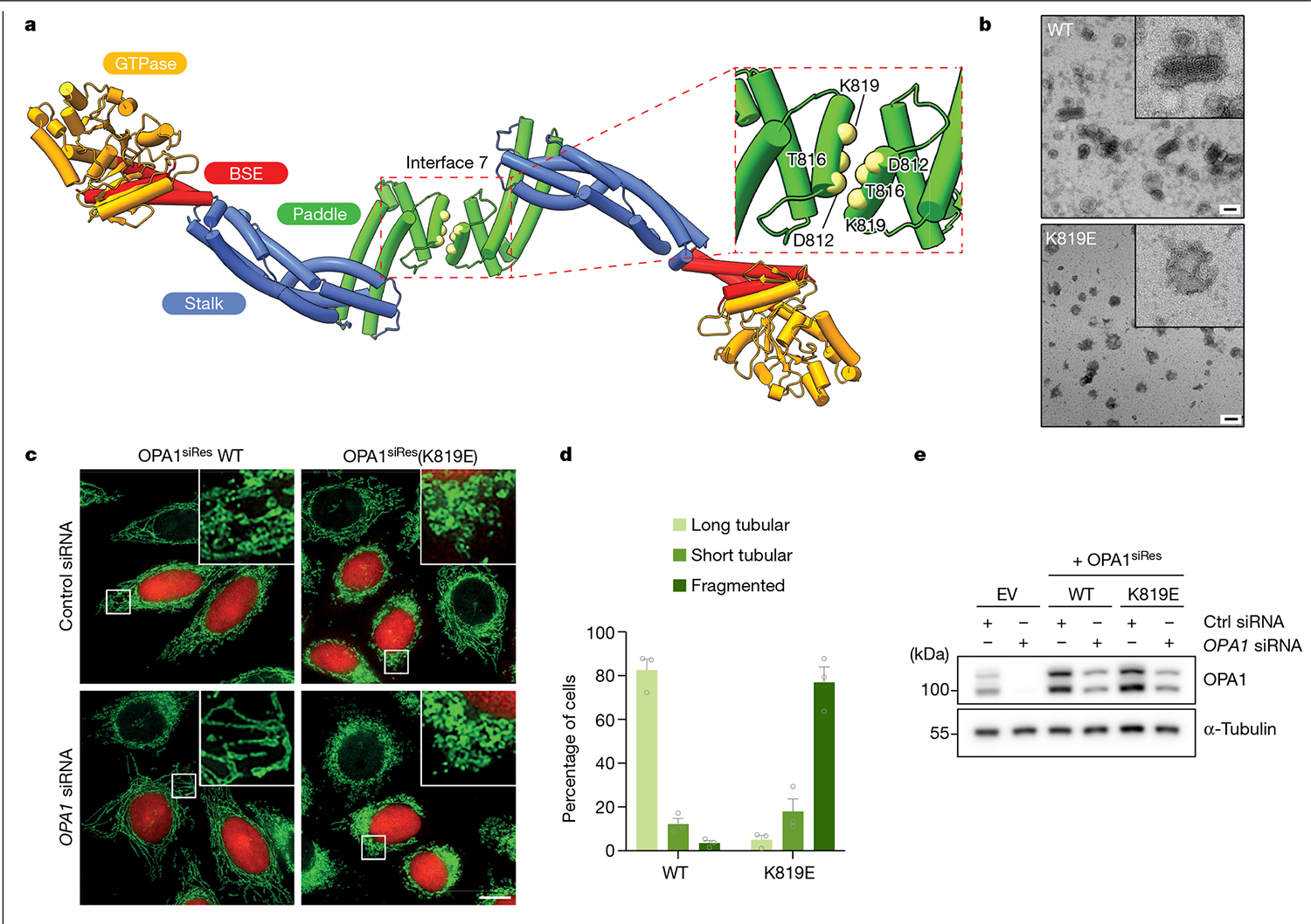
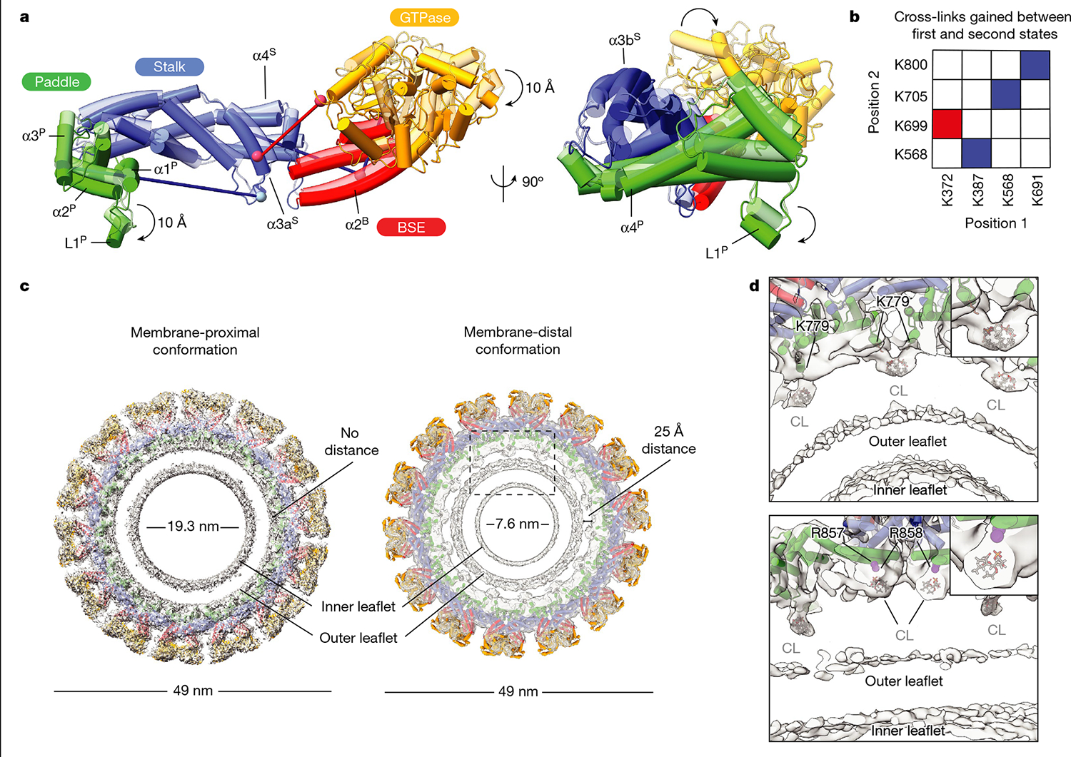
Distinct morphologies of the mitochondrial network support divergent metabolic and regulatory processes that determine cell function and fate. The mechanochemical GTPase optic atrophy 1 (OPA1) influences the architecture of cristae and catalyses the fusion of the mitochondrial inner membrane. Despite its fundamental importance, the molecular mechanisms by which OPA1 modulates mitochondrial morphology are unclear. Here, using a combination of cellular and structural analyses, we illuminate the molecular mechanisms that are key to OPA1-dependent membrane remodelling and fusion. Human OPA1 embeds itself into cardiolipin-containing membranes through a lipid-binding paddle domain. A conserved loop within the paddle domain inserts deeply into the bilayer, further stabilizing the interactions with cardiolipin-enriched membranes. OPA1 dimerization through the paddle domain promotes the helical assembly of a flexible OPA1 lattice on the membrane, which drives mitochondrial fusion in cells. Moreover, the membrane-bending OPA1 oligomer undergoes conformational changes that pull the membrane-inserting loop out of the outer leaflet and contribute to the mechanics of membrane remodelling. Our findings provide a structural framework for understanding how human OPA1 shapes mitochondrial morphology and show us how human disease mutations compromise OPA1 functions.

摘要

线粒体网络的不同形态支持决定细胞功能和命运的不同代谢和调节过程。机械化学 GTP 酶视神经萎缩 1(OPA1)影响嵴的结构并催化线粒体内膜的融合。尽管 OPA1 至关重要,但调节线粒体形态的分子机制尚不清楚。在这里,我们使用细胞和结构分析的组合,阐明了 OPA1 依赖性膜重塑和融合的关键分子机制。人 OPA1 通过含有脂酰甘油的膜中的脂质结合桨叶域嵌入其中。桨叶域内的保守环深入插入双层,进一步稳定了与富含心磷脂的膜的相互作用。通过桨叶域的 OPA1 二聚化促进了灵活的 OPA1 晶格在膜上的螺旋组装,从而促进细胞中的线粒体融合。此外,膜弯曲的 OPA1 寡聚体发生构象变化,将插入膜的环从外叶层中拉出,并有助于膜重塑的力学。我们的发现为理解人 OPA1 如何塑造线粒体形态提供了一个结构框架,并展示了人类疾病突变如何破坏 OPA1 的功能。

https://cdn.ncbi.nlm.nih.gov/pmc/blobs/5d71/10875962/1fd5f8f452f8/nihms-1962756-f0005.jpg
https://cdn.ncbi.nlm.nih.gov/pmc/blobs/5d71/10875962/c757d86a51c0/nihms-1962756-f0006.jpg
https://cdn.ncbi.nlm.nih.gov/pmc/blobs/5d71/10875962/6362c1c49f26/nihms-1962756-f0007.jpg
https://cdn.ncbi.nlm.nih.gov/pmc/blobs/5d71/10875962/d19095a6218d/nihms-1962756-f0008.jpg
https://cdn.ncbi.nlm.nih.gov/pmc/blobs/5d71/10875962/c732802ac9d4/nihms-1962756-f0009.jpg
https://cdn.ncbi.nlm.nih.gov/pmc/blobs/5d71/10875962/113275be5642/nihms-1962756-f0010.jpg
https://cdn.ncbi.nlm.nih.gov/pmc/blobs/5d71/10875962/07c761f593e9/nihms-1962756-f0011.jpg
https://cdn.ncbi.nlm.nih.gov/pmc/blobs/5d71/10875962/c5a4feda124a/nihms-1962756-f0012.jpg
https://cdn.ncbi.nlm.nih.gov/pmc/blobs/5d71/10875962/1816b2b6c89f/nihms-1962756-f0013.jpg
https://cdn.ncbi.nlm.nih.gov/pmc/blobs/5d71/10875962/d4bbd4992fa9/nihms-1962756-f0014.jpg
https://cdn.ncbi.nlm.nih.gov/pmc/blobs/5d71/10875962/fbf37ed34c2a/nihms-1962756-f0015.jpg
https://cdn.ncbi.nlm.nih.gov/pmc/blobs/5d71/10875962/412f31199728/nihms-1962756-f0001.jpg
https://cdn.ncbi.nlm.nih.gov/pmc/blobs/5d71/10875962/227e21041104/nihms-1962756-f0002.jpg
https://cdn.ncbi.nlm.nih.gov/pmc/blobs/5d71/10875962/e620e5880190/nihms-1962756-f0003.jpg
https://cdn.ncbi.nlm.nih.gov/pmc/blobs/5d71/10875962/6ff379e7b651/nihms-1962756-f0004.jpg
https://cdn.ncbi.nlm.nih.gov/pmc/blobs/5d71/10875962/1fd5f8f452f8/nihms-1962756-f0005.jpg

相似文献

1
Structural mechanism of mitochondrial membrane remodelling by human OPA1.人源 OPA1 调控线粒体膜重塑的结构机制
Nature. 2023 Aug;620(7976):1101-1108. doi: 10.1038/s41586-023-06441-6. Epub 2023 Aug 23.
2
OPA1 helical structures give perspective to mitochondrial dysfunction.OPA1 螺旋结构为线粒体功能障碍提供了新视角。
Nature. 2023 Aug;620(7976):1109-1116. doi: 10.1038/s41586-023-06462-1. Epub 2023 Aug 23.
3
Molecular basis of selective mitochondrial fusion by heterotypic action between OPA1 and cardiolipin.OPA1 与心磷脂异质作用介导的选择性线粒体融合的分子基础。
Nat Cell Biol. 2017 Jul;19(7):856-863. doi: 10.1038/ncb3560. Epub 2017 Jun 19.
4
MFN1 structures reveal nucleotide-triggered dimerization critical for mitochondrial fusion.MFN1结构揭示了核苷酸触发的二聚化对线粒体融合至关重要。
Nature. 2017 Feb 16;542(7641):372-376. doi: 10.1038/nature21077. Epub 2017 Jan 23.
5
Structure and assembly of the mitochondrial membrane remodelling GTPase Mgm1.线粒体膜重塑 GTP 酶 Mgm1 的结构与组装
Nature. 2019 Jul;571(7765):429-433. doi: 10.1038/s41586-019-1372-3. Epub 2019 Jul 10.
6
OPA1-dependent cristae modulation is essential for cellular adaptation to metabolic demand.OPA1 依赖的嵴调节对于细胞适应代谢需求至关重要。
EMBO J. 2014 Nov 18;33(22):2676-91. doi: 10.15252/embj.201488349. Epub 2014 Oct 8.
7
Relationship between OPA1 and cardiolipin in mitochondrial inner-membrane fusion.OPA1 与线粒体内膜融合中心的 cardiolipin 之间的关系。
Biochim Biophys Acta Bioenerg. 2018 Sep;1859(9):951-957. doi: 10.1016/j.bbabio.2018.05.016. Epub 2018 May 28.
8
disease-causing mutants have domain-specific effects on mitochondrial ultrastructure and fusion.致病突变对线粒体超微结构和融合有特定的影响。
Proc Natl Acad Sci U S A. 2023 Mar 21;120(12):e2207471120. doi: 10.1073/pnas.2207471120. Epub 2023 Mar 16.
9
Two forms of Opa1 cooperate to complete fusion of the mitochondrial inner-membrane.两种形式的 Opa1 协同作用完成线粒体内膜的融合。
Elife. 2020 Jan 10;9:e50973. doi: 10.7554/eLife.50973.
10
L-OPA1 regulates mitoflash biogenesis independently from membrane fusion.L-OPA1独立于膜融合调节线粒体闪光的产生。
EMBO Rep. 2017 Mar;18(3):451-463. doi: 10.15252/embr.201642931. Epub 2017 Feb 7.

引用本文的文献

1
Endothelial mitochondria in the blood-brain barrier.血脑屏障中的内皮线粒体。
Fluids Barriers CNS. 2025 Aug 25;22(1):88. doi: 10.1186/s12987-025-00699-w.
2
GLP-1R associates with VAPB and SPHKAP at ERMCSs to regulate β-cell mitochondrial remodelling and function.胰高血糖素样肽-1受体(GLP-1R)在内质网-线粒体接触位点(ERMCSs)与VAPB和SPHKAP结合,以调节β细胞线粒体重塑和功能。
bioRxiv. 2025 Jul 20:2024.04.28.591531. doi: 10.1101/2024.04.28.591531.
3
Technological advances in the diagnosis and management of inherited optic neuropathies.

本文引用的文献

1
Determinants and outcomes of mitochondrial dynamics.线粒体动力学的决定因素及结果
Mol Cell. 2023 Mar 16;83(6):857-876. doi: 10.1016/j.molcel.2023.02.012. Epub 2023 Mar 7.
2
The structure of the human LACTB filament reveals the mechanisms of assembly and membrane binding.人源 LACTB 丝状体结构揭示了其组装和膜结合的机制。
PLoS Biol. 2022 Dec 19;20(12):e3001899. doi: 10.1371/journal.pbio.3001899. eCollection 2022 Dec.
3
Structure and functionality of a multimeric human COQ7:COQ9 complex.多聚体人 COQ7:COQ9 复合物的结构与功能。
遗传性视神经病变诊断与管理的技术进展
Front Neurol. 2025 Jul 25;16:1609033. doi: 10.3389/fneur.2025.1609033. eCollection 2025.
4
PTPN2 Inhibition Disrupts Mitochondrial Renewal and Blocks TFRC-Mediated Mitophagy to Exert Anti-Tumor Activities in ALK-Positive Anaplastic Large Cell Lymphoma.蛋白酪氨酸磷酸酶非受体型2(PTPN2)抑制作用破坏线粒体更新并阻断转铁蛋白受体(TFRC)介导的线粒体自噬,从而在ALK阳性间变性大细胞淋巴瘤中发挥抗肿瘤活性。
Adv Sci (Weinh). 2025 Aug;12(31):e14282. doi: 10.1002/advs.202414282. Epub 2025 Jul 30.
5
Perspectives on mitochondrial dysfunction in the regeneration of aging skeletal muscle.衰老骨骼肌再生中线粒体功能障碍的研究视角
Cell Mol Biol Lett. 2025 Jul 28;30(1):94. doi: 10.1186/s11658-025-00771-1.
6
An integrative modelling approach to the mitochondrial cristae.一种针对线粒体嵴的整合建模方法。
Commun Biol. 2025 Jul 1;8(1):972. doi: 10.1038/s42003-025-08381-5.
7
Mitochondrial DNA depletion syndrome and its cardiac complication.线粒体DNA耗竭综合征及其心脏并发症。
Front Cardiovasc Med. 2025 Jun 10;12:1582219. doi: 10.3389/fcvm.2025.1582219. eCollection 2025.
8
The Mitochondrial Foundations of Parkinson's Disease: Therapeutic Implications.帕金森病的线粒体基础:治疗意义
Aging Dis. 2025 Apr 28;16(5):2695-2720. doi: 10.14336/AD.2025.0440.
9
Mitochondria in oxidative stress, inflammation and aging: from mechanisms to therapeutic advances.氧化应激、炎症与衰老中的线粒体:从机制到治疗进展
Signal Transduct Target Ther. 2025 Jun 11;10(1):190. doi: 10.1038/s41392-025-02253-4.
10
ER-mitochondria tethering and its signaling: A novel therapeutic target in breast cancer.内质网-线粒体连接及其信号传导:乳腺癌中的一个新治疗靶点。
Mol Ther Oncol. 2025 May 14;33(2):200995. doi: 10.1016/j.omton.2025.200995. eCollection 2025 Jun 18.
Mol Cell. 2022 Nov 17;82(22):4307-4323.e10. doi: 10.1016/j.molcel.2022.10.003. Epub 2022 Oct 27.
4
Mitochondrial cristae-remodeling protein OPA1 in POMC neurons couples Ca homeostasis with adipose tissue lipolysis.POMC 神经元中线粒体嵴重塑蛋白 OPA1 将钙稳态与脂肪组织脂解相偶联。
Cell Metab. 2021 Sep 7;33(9):1820-1835.e9. doi: 10.1016/j.cmet.2021.07.008. Epub 2021 Aug 2.
5
MitoSegNet: Easy-to-use Deep Learning Segmentation for Analyzing Mitochondrial Morphology.线粒体分割网络(MitoSegNet):用于分析线粒体形态的易于使用的深度学习分割方法。
iScience. 2020 Sep 29;23(10):101601. doi: 10.1016/j.isci.2020.101601. eCollection 2020 Oct 23.
6
UCSF ChimeraX: Structure visualization for researchers, educators, and developers.UCSF ChimeraX:面向研究人员、教育工作者和开发者的结构可视化工具。
Protein Sci. 2021 Jan;30(1):70-82. doi: 10.1002/pro.3943. Epub 2020 Oct 22.
7
Structural insights into G domain dimerization and pathogenic mutation of OPA1.OPA1 构象结构域二聚化及致病性突变的结构研究
J Cell Biol. 2020 Jul 6;219(7). doi: 10.1083/jcb.201907098.
8
CHCHD10-regulated OPA1-mitofilin complex mediates TDP-43-induced mitochondrial phenotypes associated with frontotemporal dementia.CHCHD10 调节的 OPA1-肌联蛋白复合物介导 TDP-43 诱导的与额颞叶痴呆相关的线粒体表型。
FASEB J. 2020 Jun;34(6):8493-8509. doi: 10.1096/fj.201903133RR. Epub 2020 May 5.
9
Developmental and Tumor Angiogenesis Requires the Mitochondria-Shaping Protein Opa1.发育和肿瘤血管生成需要线粒体塑形蛋白 Opa1。
Cell Metab. 2020 May 5;31(5):987-1003.e8. doi: 10.1016/j.cmet.2020.04.007. Epub 2020 Apr 20.
10
Cryo-EM structures of S-OPA1 reveal its interactions with membrane and changes upon nucleotide binding.冷冻电镜结构研究揭示 S-OPA1 与膜的相互作用及其在核苷酸结合时的构象变化。
Elife. 2020 Mar 31;9:e50294. doi: 10.7554/eLife.50294.