Khan Muhammad Shahid, Buzdar Saeed Ahmad, Hussain Riaz, Alouffi Abdulaziz, Aleem Muhammad Tahir, Farhab Muhammad, Javid Muhammad Arshad, Akhtar Rana Waseem, Khan Iahtasham, Almutairi Mashal M
Institute of Physics, The Islamia University, Bahawalpur 63100, Pakistan.
Department of Pathology, Faculty of Veterinary and Animal Sciences, The Islamia University, Bahawalpur 63100, Pakistan.
Vet Sci. 2023 Aug 9;10(8):514. doi: 10.3390/vetsci10080514.
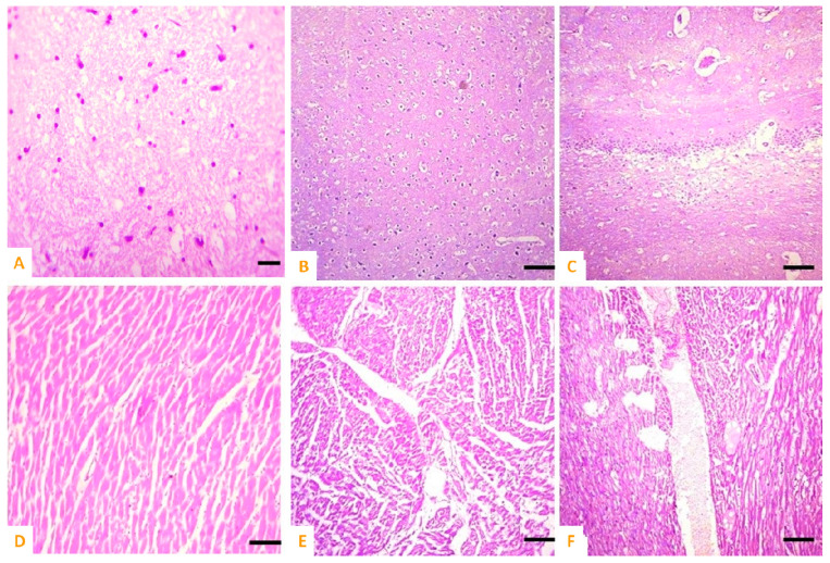
The market for nanoparticles has grown significantly over the past few decades due to a number of unique qualities, including antibacterial capabilities. It is still unclear how nanoparticle toxicity works. In order to ascertain the toxicity of synthetic cobalt iron oxide (CoFeO) nanoparticles (CIONPs) in rabbits, this study was carried out. Sixteen rabbits in total were purchased from the neighborhood market and divided into two groups (A and B), each of which contained eight rabbits. The CIONPs were synthesized by the co-precipitation method. Crystallinity and phase identification were confirmed by X-ray diffraction (XRD). The average size of the nanoparticles (13.2 nm) was calculated by Scherrer formula (Dhkl = 0.9 λ/β cos θ) and confirmed by TEM images. The saturation magnetization, 50.1 emug, was measured by vibrating sample magnetometer (VSM). CIONPs were investigated as contrast agents (CA) for magnetic resonance images (MRI). The relaxivity (r = 1/T) of the MRI was also investigated at a field strength of 0.35 T (Tesla), and the ratio r/r for the CIONPs contrast agent was 6.63. The CIONPs were administrated intravenously into the rabbits through the ear vein. Blood was collected at days 5 and 10 post-exposure for hematological and serum biochemistry analyses. The intensities of the signal experienced by CA with CIONPs were 1427 for the liver and 1702 for the spleen. The treated group showed significantly lower hematological parameters, but significantly higher total white blood cell counts and neutrophils. The results of the serum biochemistry analyses showed significantly higher and lower quantities of different serum biochemical parameters in the treated rabbits at day 10 of the trial. At the microscopic level, different histological ailments were observed in the visceral organs of treated rabbits, including the liver, kidneys, spleen, heart, and brain. In conclusion, the results revealed that cobalt iron oxide (CoFeO) nanoparticles induced toxicity via alterations in multiple tissues of rabbits.
在过去几十年中,由于包括抗菌能力在内的许多独特特性,纳米颗粒市场显著增长。纳米颗粒毒性的作用机制仍不清楚。为了确定合成钴铁氧化物(CoFeO)纳米颗粒(CIONPs)对兔子的毒性,开展了本研究。总共从附近市场购买了16只兔子,并将其分为两组(A组和B组),每组包含8只兔子。通过共沉淀法合成CIONPs。通过X射线衍射(XRD)确认结晶度和相鉴定。通过谢乐公式(Dhkl = 0.9λ/βcosθ)计算纳米颗粒的平均尺寸(13.2nm),并通过透射电子显微镜(TEM)图像进行确认。通过振动样品磁强计(VSM)测量饱和磁化强度为50.1 emug。研究了CIONPs作为磁共振成像(MRI)的造影剂(CA)。还在0.35特斯拉(T)的场强下研究了MRI的弛豫率(r = 1/T),CIONPs造影剂的r/r比率为6.63。通过耳静脉将CIONPs静脉注射到兔子体内。在暴露后第5天和第10天采集血液进行血液学和血清生化分析。含有CIONPs的CA在肝脏和脾脏处的信号强度分别为1427和1702。治疗组的血液学参数显著降低,但总白细胞计数和中性粒细胞显著升高。血清生化分析结果显示,在试验第10天,治疗组兔子的不同血清生化参数数量显著升高和降低。在显微镜水平上,在治疗组兔子的内脏器官(包括肝脏、肾脏、脾脏、心脏和大脑)中观察到不同的组织病理学病变。总之,结果表明钴铁氧化物(CoFeO)纳米颗粒通过改变兔子的多个组织诱导毒性。