Immunity and Pathogen Unit, Microbiology and Infectious Diseases Department, Institut de Recherche Biomédicale des Armées (IRBA), Brétigny-sur-Orge, France.
Respiratory Department, Percy Military Teaching Hospital, Clamart, France.
Front Immunol. 2023 Aug 15;14:1241323. doi: 10.3389/fimmu.2023.1241323. eCollection 2023.
Inflammatory lesions after Influenza A viruses (IAV) are potential therapeutic target for which better understanding of post-infection immune mechanisms is required. Most studies to evaluate innate immune reactions induced by IAV are based on quantitative/functional methods and anatomical exploration is most often non-existent. We aimed to study pulmonary damage and macrophage recruitment using two-photon excitation microscopy (TPEM) after IAV infection.
We infected C57BL/6 CD11cYFP mice with A/Puerto Ricco/8/34 H1N1. We performed immune cell analysis, including flow cytometry, cytokine concentration assays, and TPEM observations after staining with anti-F4/80 antibody coupled to BV421. We adapted live lung slice (LLS) method for intravital microscopy to analyze cell motility.
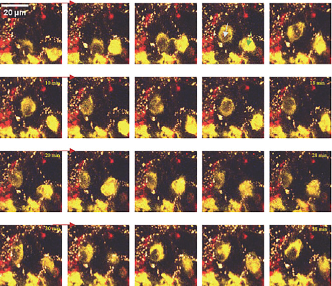
TPEM provided complementary data to flow cytometry and cytokine assays by allowing observation of bronchial epithelium lesions and spreading of local infection. Addition of F4/80-BV421 staining allowed us to precisely determine timing of recruitment and pulmonary migration of macrophages. LLS preserved cellular viability, allowing us to observe acceleration of macrophage motility.
After IAV infection, we were able to explore structural consequences and successive waves of innate immune cell recruitment. By combining microscopy, flow cytometry and chemokine measurements, we describe novel and precise scenario of innate immune response against IAV.
流感病毒(IAV)感染后的炎症病变是潜在的治疗靶点,需要更好地了解感染后免疫机制。大多数评估 IAV 诱导的固有免疫反应的研究都基于定量/功能方法,而解剖探索通常不存在。我们旨在使用双光子激发显微镜(TPEM)研究 IAV 感染后的肺部损伤和巨噬细胞募集。
我们用 A/Puerto Ricco/8/34 H1N1 感染 C57BL/6 CD11cYFP 小鼠。我们进行了免疫细胞分析,包括流式细胞术、细胞因子浓度测定和用抗 F4/80 抗体结合 BV421 进行的 TPEM 观察。我们通过活体肺切片(LLS)方法进行了活体显微镜分析,以分析细胞迁移。
TPEM 通过观察支气管上皮损伤和局部感染的扩散,为流式细胞术和细胞因子测定提供了补充数据。添加 F4/80-BV421 染色使我们能够精确确定巨噬细胞募集和肺部迁移的时间。LLS 保持了细胞活力,使我们能够观察到巨噬细胞迁移的加速。
在 IAV 感染后,我们能够探索结构后果和固有免疫细胞募集的连续波。通过结合显微镜、流式细胞术和趋化因子测量,我们描述了针对 IAV 的固有免疫反应的新的、精确的情景。