Engelke Klaus, Chaudry Oliver, Gast Lena, Eldib Mootaz Ab, Wang Ling, Laredo Jean-Denis, Schett Georg, Nagel Armin M
Department of Medicine III, Friedrich-Alexander University of Erlangen-Nürnberg, University Hospital Erlangen, Ulmenweg 18, 91054, Erlangen, Germany.
Institute of Medical Physics (IMP), Friedrich-Alexander-Universität Erlangen-Nürnberg (FAU), Henkestr. 91, 91052, Erlangen, Germany.
J Orthop Translat. 2023 Aug 19;42:57-72. doi: 10.1016/j.jot.2023.07.005. eCollection 2023 Sep.
Magnetic resonance imaging (MRI) is the dominant 3D imaging modality to quantify muscle properties in skeletal muscle disorders, in inherited and acquired muscle diseases, and in sarcopenia, in cachexia and frailty.
This review covers T1 weighted and Dixon sequences, introduces T2 mapping, diffusion tensor imaging (DTI) and non-proton MRI. Technical concepts, strengths, limitations and translational aspects of these techniques are discussed in detail. Examples of clinical applications are outlined. For comparison P-and C-MR Spectroscopy are also addressed.
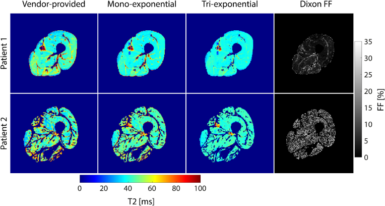
MRI technology provides a rich toolset to assess muscle deterioration. In addition to classical measures such as muscle atrophy using T1 weighted imaging and fat infiltration using Dixon sequences, parameters characterizing inflammation from T2 maps, tissue sodium using non-proton MRI techniques or concentration or fiber architecture using diffusion tensor imaging may be useful for an even earlier diagnosis of the impairment of muscle quality.
Quantitative MRI provides new options for muscle research and clinical applications. Current limitations that also impair its more widespread use in clinical trials are lack of standardization, ambiguity of image segmentation and analysis approaches, a multitude of outcome parameters without a clear strategy which ones to use and the lack of normal data.
磁共振成像(MRI)是用于量化骨骼肌疾病、遗传性和获得性肌肉疾病、肌肉减少症、恶病质和虚弱症中肌肉特性的主要三维成像方式。
本综述涵盖T1加权和狄克逊序列,介绍T2 mapping、扩散张量成像(DTI)和非质子MRI。详细讨论了这些技术的技术概念、优势、局限性及转化方面。概述了临床应用实例。为作比较,还讨论了磷磁共振波谱和碳磁共振波谱。
MRI技术提供了一套丰富的工具来评估肌肉退化。除了使用T1加权成像测量肌肉萎缩和使用狄克逊序列测量脂肪浸润等经典方法外,通过T2 mapping表征炎症的参数、使用非质子MRI技术测量组织钠含量或使用扩散张量成像测量浓度或纤维结构,可能有助于更早诊断肌肉质量受损情况。
定量MRI为肌肉研究和临床应用提供了新的选择。目前阻碍其在临床试验中更广泛应用的局限性包括缺乏标准化、图像分割和分析方法不明确、众多结局参数且没有明确使用哪些参数的策略以及缺乏正常数据。