Qian Hongwei, Shen Zhihong, Zhou Difan, Huang Yanhua
Department of Hepatobiliary and Pancreatic Surgery, Shaoxing People's Hospital, Shaoxing, China.
Shaoxing Key Laboratory of Minimally Invasive Abdominal Surgery and Precise Treatment of Tumor, Shaoxing, China.
Front Oncol. 2023 Aug 24;13:1209111. doi: 10.3389/fonc.2023.1209111. eCollection 2023.
Hepatocellular cancer (HCC) is one of the most common tumors worldwide, and Ki-67 is highly important in the assessment of HCC. Our study aimed to evaluate the value of ultrasound radiomics based on intratumoral and peritumoral tissues in predicting Ki-67 expression levels in patients with HCC.
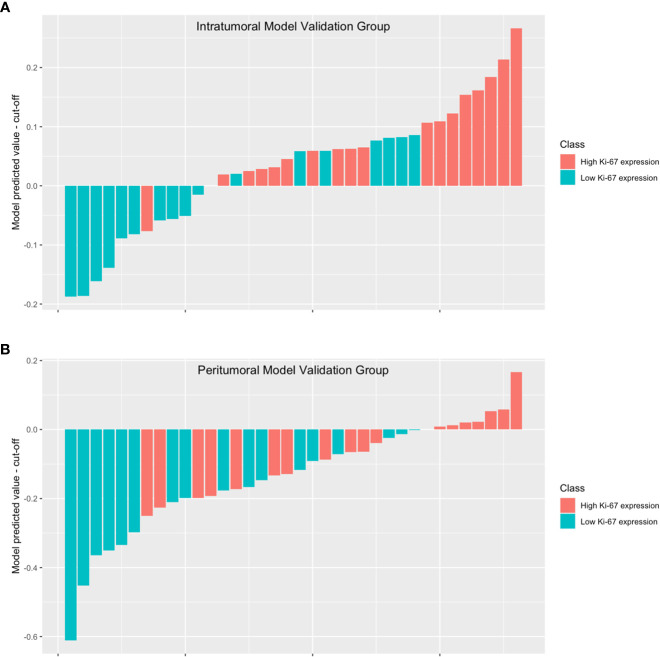
We conducted a retrospective analysis of ultrasonic and clinical data from 118 patients diagnosed with HCC through histopathological examination of surgical specimens in our hospital between September 2019 and January 2023. Radiomics features were extracted from ultrasound images of both intratumoral and peritumoral regions. To select the optimal features, we utilized the t-test and the least absolute shrinkage and selection operator (LASSO). We compared the area under the curve (AUC) values to determine the most effective modeling method. Subsequently, we developed four models: the intratumoral model, the peritumoral model, combined model #1, and combined model #2.
Of the 118 patients, 64 were confirmed to have high Ki-67 expression while 54 were confirmed to have low Ki-67 expression. The AUC of the intratumoral model was 0.796 (0.649-0.942), and the AUC of the peritumoral model was 0.772 (0.619-0.926). Furthermore, combined model#1 yielded an AUC of 0.870 (0.751-0.989), and the AUC of combined model#2 was 0.762 (0.605-0.918). Among these models, combined model#1 showed the best performance in terms of AUC, accuracy, F1-score, and decision curve analysis (DCA).
We presented an ultrasound radiomics model that utilizes both intratumoral and peritumoral tissue information to accurately predict Ki-67 expression in HCC patients. We believe that incorporating both regions in a proper manner can enhance the diagnostic performance of the prediction model. Nevertheless, it is not sufficient to include both regions in the region of interest (ROI) without careful consideration.
肝细胞癌(HCC)是全球最常见的肿瘤之一,Ki-67在HCC评估中非常重要。本研究旨在评估基于肿瘤内和瘤周组织的超声放射组学在预测HCC患者Ki-67表达水平方面的价值。
我们对2019年9月至2023年1月期间在我院通过手术标本组织病理学检查确诊为HCC的118例患者的超声和临床数据进行了回顾性分析。从肿瘤内和瘤周区域的超声图像中提取放射组学特征。为了选择最佳特征,我们使用了t检验和最小绝对收缩和选择算子(LASSO)。我们比较曲线下面积(AUC)值以确定最有效的建模方法。随后,我们开发了四个模型:肿瘤内模型、瘤周模型、联合模型#1和联合模型#2。
118例患者中,64例确诊为Ki-67高表达,54例确诊为Ki-67低表达。肿瘤内模型的AUC为0.796(0.649 - 0.942),瘤周模型的AUC为0.772(0.619 - 0.926)。此外,联合模型#1的AUC为0.870(0.751 - 0.989),联合模型#2的AUC为O.762(0.605 - 0.918)。在这些模型中,联合模型#1在AUC、准确性、F1分数和决策曲线分析(DCA)方面表现最佳。
我们提出了一种超声放射组学模型,该模型利用肿瘤内和瘤周组织信息准确预测HCC患者的Ki-67表达。我们认为以适当方式纳入这两个区域可以提高预测模型的诊断性能。然而,在没有仔细考虑的情况下仅将这两个区域纳入感兴趣区域(ROI)是不够的。