• 文献检索
  • 文档翻译
  • 深度研究
  • 学术资讯
  • Suppr Zotero 插件Zotero 插件
  • 邀请有礼
  • 套餐&价格
  • 历史记录
应用&插件
Suppr Zotero 插件Zotero 插件浏览器插件Mac 客户端Windows 客户端微信小程序
定价
高级版会员购买积分包购买API积分包
服务
文献检索文档翻译深度研究API 文档MCP 服务
关于我们
关于 Suppr公司介绍联系我们用户协议隐私条款
关注我们

Suppr 超能文献

核心技术专利:CN118964589B侵权必究
粤ICP备2023148730 号-1Suppr @ 2026

文献检索

告别复杂PubMed语法,用中文像聊天一样搜索,搜遍4000万医学文献。AI智能推荐,让科研检索更轻松。

立即免费搜索

文件翻译

保留排版,准确专业,支持PDF/Word/PPT等文件格式,支持 12+语言互译。

免费翻译文档

深度研究

AI帮你快速写综述,25分钟生成高质量综述,智能提取关键信息,辅助科研写作。

立即免费体验

基于深度学习的肺结节检测系统对肺结节位置影响的诊断性能及临床价值

The diagnostic performance and clinical value of deep learning-based nodule detection system concerning influence of location of pulmonary nodule.

作者信息

You Seulgi, Park Ji Hyun, Park Bumhee, Shin Han-Bit, Ha Taeyang, Yun Jae Sung, Park Kyoung Joo, Jung Yongjun, Kim You Na, Kim Minji, Sun Joo Sung

机构信息

Department of Radiology, Ajou University School of Medicine, Suwon, Republic of Korea.

Office of Biostatistics, Ajou Research Institute for Innovative Medicine, Ajou University Medical Center, Suwon, Republic of Korea.

出版信息

Insights Imaging. 2023 Sep 19;14(1):149. doi: 10.1186/s13244-023-01497-4.

DOI:10.1186/s13244-023-01497-4
PMID:37726452
原文链接:https://pmc.ncbi.nlm.nih.gov/articles/PMC10509107/
Abstract

BACKGROUND

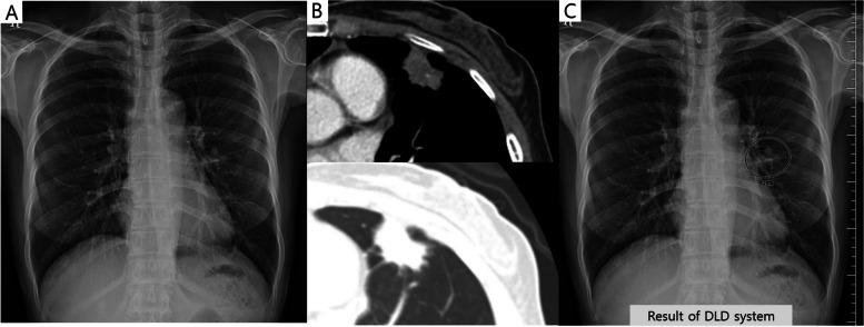
The deep learning-based nodule detection (DLD) system improves nodule detection performance of observers on chest radiographs (CXRs). However, its performance in different pulmonary nodule (PN) locations remains unknown.

METHODS

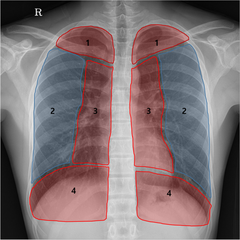
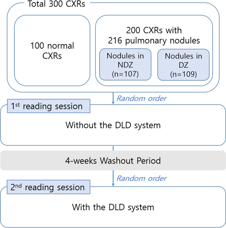
We divided the CXR intrathoracic region into non-danger zone (NDZ) and danger zone (DZ). The DZ included the lung apices, paramediastinal areas, and retrodiaphragmatic areas, where nodules could be missed. We used a dataset of 300 CXRs (100 normal and 200 abnormal images with 216 PNs [107 NDZ and 109 DZ nodules]). Eight observers (two thoracic radiologists [TRs], two non-thoracic radiologists [NTRs], and four radiology residents [RRs]) interpreted each radiograph with and without the DLD system. The metric of lesion localization fraction (LLF; the number of correctly localized lesions divided by the total number of true lesions) was used to evaluate the diagnostic performance according to the nodule location.

RESULTS

The DLD system demonstrated a lower LLF for the detection of DZ nodules (64.2) than that of NDZ nodules (83.2, p = 0.008). For DZ nodule detection, the LLF of the DLD system (64.2) was lower than that of TRs (81.7, p < 0.001), which was comparable to that of NTRs (56.4, p = 0.531) and RRs (56.7, p = 0.459). Nonetheless, the LLF of RRs significantly improved from 56.7 to 65.6 using the DLD system (p = 0.021) for DZ nodule detection.

CONCLUSION

The performance of the DLD system was lower in the detection of DZ nodules compared to that of NDZ nodules. Nonetheless, RR performance in detecting DZ nodules improved upon using the DLD system.

CRITICAL RELEVANCE STATEMENT

Despite the deep learning-based nodule detection system's limitations in detecting danger zone nodules, it proves beneficial for less-experienced observers by providing valuable assistance in identifying these nodules, thereby advancing nodule detection in clinical practice.

KEY POINTS

• The deep learning-based nodule detection (DLD) system can improve the diagnostic performance of observers in nodule detection. • The DLD system shows poor diagnostic performance in detecting danger zone nodules. • For less-experienced observers, the DLD system is helpful in detecting danger zone nodules.

摘要

背景

基于深度学习的结节检测(DLD)系统提高了观察者对胸部X光片(CXR)的结节检测性能。然而,其在不同肺结节(PN)位置的性能仍不清楚。

方法

我们将CXR胸腔内区域分为非危险区(NDZ)和危险区(DZ)。DZ包括肺尖、纵隔旁区域和膈后区域,这些区域的结节可能会被漏诊。我们使用了一个包含300张CXR的数据集(100张正常图像和200张异常图像,有216个PN[107个NDZ结节和109个DZ结节])。八名观察者(两名胸科放射科医生[TRs]、两名非胸科放射科医生[NTRs]和四名放射科住院医师[RRs])在有和没有DLD系统的情况下解读每张X光片。使用病变定位分数(LLF;正确定位的病变数量除以真实病变总数)指标根据结节位置评估诊断性能。

结果

DLD系统检测DZ结节的LLF(64.2)低于NDZ结节(83.2,p = 0.008)。对于DZ结节检测,DLD系统的LLF(64.2)低于TRs(81.7,p < 0.001),与NTRs(56.4,p = 0.531)和RRs(56.7,p = 0.459)相当。尽管如此,对于DZ结节检测,使用DLD系统时RRs的LLF从56.7显著提高到65.6(p = 0.021)。

结论

与NDZ结节相比,DLD系统在检测DZ结节方面的性能较低。尽管如此,RRs在使用DLD系统检测DZ结节时的性能有所提高。

关键相关性声明

尽管基于深度学习的结节检测系统在检测危险区结节方面存在局限性,但它通过在识别这些结节方面提供有价值的帮助,证明对经验较少的观察者有益,从而推动了临床实践中的结节检测。

要点

• 基于深度学习的结节检测(DLD)系统可以提高观察者在结节检测中的诊断性能。• DLD系统在检测危险区结节方面显示出较差的诊断性能。• 对于经验较少的观察者,DLD系统有助于检测危险区结节。

https://cdn.ncbi.nlm.nih.gov/pmc/blobs/e2bd/10509107/9a61f48764f9/13244_2023_1497_Fig5_HTML.jpg
https://cdn.ncbi.nlm.nih.gov/pmc/blobs/e2bd/10509107/8d111ab7998f/13244_2023_1497_Fig1_HTML.jpg
https://cdn.ncbi.nlm.nih.gov/pmc/blobs/e2bd/10509107/8f6edff68477/13244_2023_1497_Fig2_HTML.jpg
https://cdn.ncbi.nlm.nih.gov/pmc/blobs/e2bd/10509107/f8495a8f9cb5/13244_2023_1497_Fig3_HTML.jpg
https://cdn.ncbi.nlm.nih.gov/pmc/blobs/e2bd/10509107/29f7f3de7226/13244_2023_1497_Fig4_HTML.jpg
https://cdn.ncbi.nlm.nih.gov/pmc/blobs/e2bd/10509107/9a61f48764f9/13244_2023_1497_Fig5_HTML.jpg

相似文献

1
The diagnostic performance and clinical value of deep learning-based nodule detection system concerning influence of location of pulmonary nodule.基于深度学习的肺结节检测系统对肺结节位置影响的诊断性能及临床价值
Insights Imaging. 2023 Sep 19;14(1):149. doi: 10.1186/s13244-023-01497-4.
2
Deep learning-based detection system for multiclass lesions on chest radiographs: comparison with observer readings.基于深度学习的胸片多类病变检测系统:与观察者读数的比较。
Eur Radiol. 2020 Mar;30(3):1359-1368. doi: 10.1007/s00330-019-06532-x. Epub 2019 Nov 20.
3
The advantage of digital tomosynthesis for pulmonary nodule detection concerning influence of nodule location and size: a phantom study.数字断层合成技术在肺结节检测中关于结节位置和大小影响的优势:一项体模研究
Clin Radiol. 2017 Sep;72(9):796.e1-796.e8. doi: 10.1016/j.crad.2017.03.022. Epub 2017 Apr 25.
4
Bone Suppression on Chest Radiographs for Pulmonary Nodule Detection: Comparison between a Generative Adversarial Network and Dual-Energy Subtraction.胸部 X 线摄影检测肺结节时的骨抑制:生成对抗网络与双能减影的比较。
Korean J Radiol. 2022 Jan;23(1):139-149. doi: 10.3348/kjr.2021.0146.
5
Added Value of Deep Learning-based Detection System for Multiple Major Findings on Chest Radiographs: A Randomized Crossover Study.深度学习检测系统对胸部 X 线片中多个主要发现的增值作用:一项随机交叉研究。
Radiology. 2021 May;299(2):450-459. doi: 10.1148/radiol.2021202818. Epub 2021 Mar 23.
6
Validation of a Deep Learning Algorithm for the Detection of Malignant Pulmonary Nodules in Chest Radiographs.深度学习算法在胸部 X 光片中检测恶性肺结节的验证。
JAMA Netw Open. 2020 Sep 1;3(9):e2017135. doi: 10.1001/jamanetworkopen.2020.17135.
7
Deep learning-based automatic detection for pulmonary nodules on chest radiographs: The relationship with background lung condition, nodule characteristics, and location.基于深度学习的胸部 X 线片肺结节自动检测:与背景肺状况、结节特征和位置的关系。
Eur J Radiol. 2023 Sep;166:111002. doi: 10.1016/j.ejrad.2023.111002. Epub 2023 Jul 22.
8
Deep Convolutional Neural Network-based Software Improves Radiologist Detection of Malignant Lung Nodules on Chest Radiographs.基于深度卷积神经网络的软件提高放射科医生在胸部 X 光片上检测恶性肺结节的能力。
Radiology. 2020 Jan;294(1):199-209. doi: 10.1148/radiol.2019182465. Epub 2019 Nov 12.
9
Performance of an AI based CAD system in solid lung nodule detection on chest phantom radiographs compared to radiology residents and fellow radiologists.基于人工智能的计算机辅助检测(CAD)系统在胸部体模X光片上检测实性肺结节的性能与放射科住院医师和放射科专科医师的比较。
J Thorac Dis. 2021 May;13(5):2728-2737. doi: 10.21037/jtd-20-3522.
10
[The performance of digital chest radiographs in the detection and diagnosis of pulmonary nodules and the consistency among readers].[数字化胸部X线片在肺结节检测与诊断中的表现及阅片者之间的一致性]
Zhonghua Zhong Liu Za Zhi. 2023 Mar 23;45(3):265-272. doi: 10.3760/cma.j.cn112152-20220304-00150.

引用本文的文献

1
Optimized deep learning model for diagnosing tonsil and adenoid hypertrophy through X-rays.通过X射线诊断扁桃体和腺样体肥大的优化深度学习模型。
Front Oncol. 2025 Mar 11;15:1508525. doi: 10.3389/fonc.2025.1508525. eCollection 2025.
2
A Systematic Review: The Role of Artificial Intelligence in Lung Cancer Screening in Detecting Lung Nodules on Chest X-Rays.一项系统评价:人工智能在肺癌筛查中对胸部X线片上肺结节检测的作用
Diagnostics (Basel). 2025 Jan 22;15(3):246. doi: 10.3390/diagnostics15030246.
3
Better performance of deep learning pulmonary nodule detection using chest radiography with pixel level labels in reference to computed tomography: data quality matters.

本文引用的文献

1
Missed Lung Cancers on Chest Radiograph: An Illustrative Review of Common Blind Spots on Chest Radiograph with Emphasis on Various Radiologic Presentations of Lung Cancers.胸部X线片漏诊的肺癌:胸部X线片常见盲点的实例综述,重点关注肺癌的各种放射学表现
Taehan Yongsang Uihakhoe Chi. 2020 Mar;81(2):351-364. doi: 10.3348/jksr.2020.81.2.351. Epub 2020 Feb 18.
2
Use of Artificial Intelligence-Based Software as Medical Devices for Chest Radiography: A Position Paper from the Korean Society of Thoracic Radiology.基于人工智能的软件作为胸部X线摄影医疗设备的应用:韩国胸放射学会立场文件
Korean J Radiol. 2021 Nov;22(11):1743-1748. doi: 10.3348/kjr.2021.0544. Epub 2021 Sep 13.
3
使用带有像素级标签的胸部 X 光片对深度学习肺结节检测的性能提升:数据质量很重要。
Sci Rep. 2024 Jul 10;14(1):15967. doi: 10.1038/s41598-024-66530-y.
4
Enhancing surgical decision-making in NEC with ResNet18: a deep learning approach to predict the need for surgery through x-ray image analysis.利用ResNet18增强坏死性小肠结肠炎的手术决策:一种通过X射线图像分析预测手术需求的深度学习方法。
Front Pediatr. 2024 Jun 4;12:1405780. doi: 10.3389/fped.2024.1405780. eCollection 2024.
Artificial intelligence in radiology: 100 commercially available products and their scientific evidence.
放射学中的人工智能:100种商用产品及其科学证据。
Eur Radiol. 2021 Jun;31(6):3797-3804. doi: 10.1007/s00330-021-07892-z. Epub 2021 Apr 15.
4
Added Value of Deep Learning-based Detection System for Multiple Major Findings on Chest Radiographs: A Randomized Crossover Study.深度学习检测系统对胸部 X 线片中多个主要发现的增值作用:一项随机交叉研究。
Radiology. 2021 May;299(2):450-459. doi: 10.1148/radiol.2021202818. Epub 2021 Mar 23.
5
Deep Learning-based Automatic Detection Algorithm for Reducing Overlooked Lung Cancers on Chest Radiographs.基于深度学习的降低胸部 X 光片漏诊肺癌的自动检测算法。
Radiology. 2020 Sep;296(3):652-661. doi: 10.1148/radiol.2020200165. Epub 2020 Jul 21.
6
Deep learning-based detection system for multiclass lesions on chest radiographs: comparison with observer readings.基于深度学习的胸片多类病变检测系统:与观察者读数的比较。
Eur Radiol. 2020 Mar;30(3):1359-1368. doi: 10.1007/s00330-019-06532-x. Epub 2019 Nov 20.
7
Development and Validation of Deep Learning-based Automatic Detection Algorithm for Malignant Pulmonary Nodules on Chest Radiographs.基于深度学习的胸部 X 线片恶性肺结节自动检测算法的开发与验证。
Radiology. 2019 Jan;290(1):218-228. doi: 10.1148/radiol.2018180237. Epub 2018 Sep 25.
8
The advantage of digital tomosynthesis for pulmonary nodule detection concerning influence of nodule location and size: a phantom study.数字断层合成技术在肺结节检测中关于结节位置和大小影响的优势:一项体模研究
Clin Radiol. 2017 Sep;72(9):796.e1-796.e8. doi: 10.1016/j.crad.2017.03.022. Epub 2017 Apr 25.
9
Pitfalls in chest radiographic interpretation: blind spots.胸部X线解读中的陷阱:盲点。
Semin Roentgenol. 2015 Jul;50(3):197-209. doi: 10.1053/j.ro.2015.01.008. Epub 2015 Feb 3.
10
The effect of computer-aided detection on radiologist performance in the detection of lung cancers previously missed on a chest radiograph.计算机辅助检测对放射科医生检测胸部 X 线片上漏诊肺癌的影响。
J Thorac Imaging. 2013 Jul;28(4):244-52. doi: 10.1097/RTI.0b013e31826c29ec.