• 文献检索
  • 文档翻译
  • 深度研究
  • 学术资讯
  • Suppr Zotero 插件Zotero 插件
  • 邀请有礼
  • 套餐&价格
  • 历史记录
应用&插件
Suppr Zotero 插件Zotero 插件浏览器插件Mac 客户端Windows 客户端微信小程序
定价
高级版会员购买积分包购买API积分包
服务
文献检索文档翻译深度研究API 文档MCP 服务
关于我们
关于 Suppr公司介绍联系我们用户协议隐私条款
关注我们

Suppr 超能文献

核心技术专利:CN118964589B侵权必究
粤ICP备2023148730 号-1Suppr @ 2026

文献检索

告别复杂PubMed语法,用中文像聊天一样搜索,搜遍4000万医学文献。AI智能推荐,让科研检索更轻松。

立即免费搜索

文件翻译

保留排版,准确专业,支持PDF/Word/PPT等文件格式,支持 12+语言互译。

免费翻译文档

深度研究

AI帮你快速写综述,25分钟生成高质量综述,智能提取关键信息,辅助科研写作。

立即免费体验

血管内超声辅助诊断和修正主动脉内椎弓根螺钉:病例说明

Intravascular ultrasound to aid in the diagnosis and revision of an intra-aortic pedicle screw: illustrative case.

作者信息

Ehlers Landon D, Opperman Patrick J, Mordeson Jack E, Thompson Jonathan R, Surdell Daniel L

机构信息

Departments of1Neurosurgery and.

2Surgery, University of Nebraska, Omaha, Nebraska.

出版信息

J Neurosurg Case Lessons. 2023 Aug 14;6(7). doi: 10.3171/CASE23272.

DOI:10.3171/CASE23272
PMID:37728279
原文链接:https://pmc.ncbi.nlm.nih.gov/articles/PMC10555651/
Abstract

BACKGROUND

Pedicle screw impingement on vessel walls has the potential for complications due to pulsatile effects and wall erosion. Artifacts from spinal instrumentation create difficulty in accurately evaluating this interface. The authors present the first case of intravascular ultrasound (IVUS) used to characterize a pedicle screw breach into the aortic lumen.

OBSERVATIONS

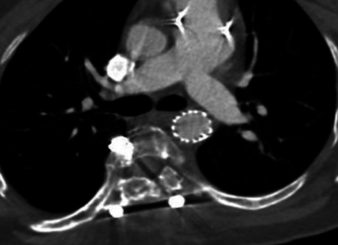
A 21-year-old female with surgically corrected scoliosis underwent computed tomography angiography (CTA) 3 years postoperatively, which revealed a pedicle screw within the thoracic aorta lumen. Metal artifact distorted the CTA images, which prompted the decision to use intraoperative IVUS. The IVUS confirmed the noninvasive imaging findings and guided final decisions regarding aortic endograft size and location during spine hardware revision.

LESSONS

For asymptomatic patients presenting with pedicle screws malpositioned in or near the aorta, treatment decisions revolve around the extent of vessel wall penetration. Intraluminal depth can be obscured by artifact on computed tomography or magnetic resonance imaging or inadequately evaluated by a transesophageal echocardiogram. In our intraoperative experience, IVUS confirmed the depth of vessel lumen violation by a single pedicle screw and no wall penetration by two additional screws of concern. This was useful in deciding on thoracic endovascular aortic repair graft size and landing zone and facilitated safe spinal instrumentation removal and revision.

摘要

背景

由于搏动效应和血管壁侵蚀,椎弓根螺钉撞击血管壁有可能引发并发症。脊柱内固定器械产生的伪影给准确评估该界面带来困难。作者报告了首例使用血管内超声(IVUS)来描述椎弓根螺钉穿破进入主动脉腔情况的病例。

观察结果

一名21岁女性,脊柱侧弯手术矫正术后3年,接受计算机断层扫描血管造影(CTA)检查,结果显示一枚椎弓根螺钉位于胸主动脉腔内。金属伪影使CTA图像失真,这促使决定术中使用IVUS。IVUS证实了无创成像结果,并在脊柱内固定器械翻修期间指导了关于主动脉内支架尺寸和位置的最终决策。

经验教训

对于主动脉内或主动脉附近椎弓根螺钉位置不当的无症状患者,治疗决策围绕血管壁穿透程度展开。计算机断层扫描或磁共振成像上的伪影可能会掩盖管腔内深度,经食管超声心动图也可能无法充分评估。根据我们的术中经验,IVUS证实了一枚椎弓根螺钉侵犯血管腔的深度,另外两枚相关螺钉未穿透血管壁。这有助于确定胸段血管腔内主动脉修复移植物的尺寸和着陆区,并便于安全地取出和翻修脊柱内固定器械。

https://cdn.ncbi.nlm.nih.gov/pmc/blobs/9134/10555651/b818476d2749/CASE23272f5.jpg
https://cdn.ncbi.nlm.nih.gov/pmc/blobs/9134/10555651/9233d01cd7ce/CASE23272f4.jpg
https://cdn.ncbi.nlm.nih.gov/pmc/blobs/9134/10555651/b818476d2749/CASE23272f5.jpg
https://cdn.ncbi.nlm.nih.gov/pmc/blobs/9134/10555651/9233d01cd7ce/CASE23272f4.jpg
https://cdn.ncbi.nlm.nih.gov/pmc/blobs/9134/10555651/b818476d2749/CASE23272f5.jpg

相似文献

1
Intravascular ultrasound to aid in the diagnosis and revision of an intra-aortic pedicle screw: illustrative case.血管内超声辅助诊断和修正主动脉内椎弓根螺钉:病例说明
J Neurosurg Case Lessons. 2023 Aug 14;6(7). doi: 10.3171/CASE23272.
2
Malpositioned pedicle screw in spine deformity surgery endangering the aorta: report of two cases, review of literature, and proposed management algorithm.脊柱畸形手术中椎弓根螺钉位置不当危及主动脉:两例报告、文献综述及拟议的处理算法
Spine Deform. 2020 Aug;8(4):809-817. doi: 10.1007/s43390-020-00094-5. Epub 2020 Mar 13.
3
Management of a periaortic spinal pedicle screw in a patient with adult presentation thoracic scoliosis.成人特发性胸段脊柱侧弯患者主动脉周围椎弓根螺钉的处理
J Orthop Case Rep. 2024 Jan;14(1):40-43. doi: 10.13107/jocr.2024.v14.i01.4140.
4
Aortic Injury by Thoracic Pedicle Screw. When Is Aortic Repair Required? Literature Review and Three New Cases.胸椎弓根螺钉致主动脉损伤。何时需要主动脉修复?文献复习及三例新病例。
World Neurosurg. 2019 Aug;128:216-224. doi: 10.1016/j.wneu.2019.04.173. Epub 2019 May 9.
5
Evaluation of pedicle screw placement in the deformed spine using intraoperative plain radiographs: a comparison with computerized tomography.使用术中平片评估畸形脊柱椎弓根螺钉置入情况:与计算机断层扫描的比较。
Spine (Phila Pa 1976). 2005 Sep 15;30(18):2084-8. doi: 10.1097/01.brs.0000178818.92105.ec.
6
Delayed open treatment of aortic penetration by a thoracic pedicle screw: illustrative case.经胸椎弓根螺钉延迟开放治疗主动脉穿透伤:病例报告
J Neurosurg Case Lessons. 2023 Feb 27;5(9). doi: 10.3171/CASE22533.
7
Management of major vascular injury during pedicle screw instrumentation of thoracolumbar spine.胸腰椎椎弓根螺钉内固定术中主要血管损伤的处理
Clin Neurol Neurosurg. 2017 Dec;163:53-59. doi: 10.1016/j.clineuro.2017.10.011. Epub 2017 Oct 16.
8
Incidence and clinical significance of vascular encroachment resulting from freehand placement of pedicle screws in the thoracic and lumbar spine: analysis of 6816 consecutive screws.经皮椎弓根螺钉置入术致胸腰椎血管侵犯的发生率及临床意义:6816枚连续螺钉的分析
Spine (Phila Pa 1976). 2014 Apr 15;39(8):683-7. doi: 10.1097/BRS.0000000000000221.
9
Complications of pedicle screw fixation in scoliosis surgery: a systematic review.脊柱侧凸手术中椎弓根螺钉固定的并发症:系统评价。
Spine (Phila Pa 1976). 2010 May 15;35(11):E465-70. doi: 10.1097/BRS.0b013e3181d1021a.
10
Intraoperative 3-dimensional imaging (O-arm) for assessment of pedicle screw position: Does it prevent unacceptable screw placement?术中三维成像(O型臂)用于评估椎弓根螺钉位置:它能否防止不可接受的螺钉置入?
Int J Spine Surg. 2012 Dec 1;6:49-54. doi: 10.1016/j.ijsp.2011.11.002. eCollection 2012.

本文引用的文献

1
Safety and Accuracy of Freehand Pedicle Screw Placement and the Role of Intraoperative O-Arm: A Single-Institution Experience.徒手椎弓根螺钉置入的安全性和准确性以及术中O型臂的作用:单机构经验
Spine (Phila Pa 1976). 2023 Feb 1;48(3):180-188. doi: 10.1097/BRS.0000000000004497. Epub 2022 Sep 28.
2
Aortic Injury by Thoracic Pedicle Screw. When Is Aortic Repair Required? Literature Review and Three New Cases.胸椎弓根螺钉致主动脉损伤。何时需要主动脉修复?文献复习及三例新病例。
World Neurosurg. 2019 Aug;128:216-224. doi: 10.1016/j.wneu.2019.04.173. Epub 2019 May 9.
3
Endovascular Treatment of Late Aortic Erosive Lesion by Pedicle Screw without Screw Removal: Case Report and Literature Review.
不带螺钉取出术的椎弓根螺钉治疗晚期主动脉侵蚀性病变:病例报告及文献综述
Ann Vasc Surg. 2017 Feb;39:285.e17-285.e21. doi: 10.1016/j.avsg.2016.06.020. Epub 2016 Sep 22.
4
Accuracy and safety of pedicle screw placement in neuromuscular scoliosis with free-hand technique.徒手技术在神经肌肉型脊柱侧凸中椎弓根螺钉置入的准确性和安全性
Eur Spine J. 2008 Dec;17(12):1686-96. doi: 10.1007/s00586-008-0795-6. Epub 2008 Oct 1.
5
Complications of thoracic pedicle screws in scoliosis treatment.脊柱侧弯治疗中胸椎椎弓根螺钉的并发症
Spine (Phila Pa 1976). 2007 Jul 1;32(15):1655-61. doi: 10.1097/BRS.0b013e318074d604.
6
Thoracic vertebral screw impingement on the aorta in an in vivo bovine model.体内牛模型中胸椎螺钉对主动脉的撞击
Spine (Phila Pa 1976). 2005 Nov 1;30(21):2406-13. doi: 10.1097/01.brs.0000184587.25330.40.
7
Vascular complications related to lumbar disc surgery.与腰椎间盘手术相关的血管并发症。
J Neurosurg. 2004 Mar;100(3 Suppl Spine):249-53. doi: 10.3171/spi.2004.100.3.0249.
8
The position of the aorta relative to the spine: a comparison of patients with and without idiopathic scoliosis.主动脉相对于脊柱的位置:特发性脊柱侧弯患者与非特发性脊柱侧弯患者的比较。
J Bone Joint Surg Am. 2003 Aug;85(8):1461-9.
9
Delayed aortic rupture caused by an implanted anterior spinal device. Case report.植入前路脊柱器械导致的延迟性主动脉破裂。病例报告。
J Neurosurg. 2001 Oct;95(2 Suppl):253-6. doi: 10.3171/spi.2001.95.2.0253.
10
False aneurysm of the thoracic aorta associated with an aorto-chest wall fistula after spinal instrumentation.脊柱内固定术后胸主动脉假性动脉瘤合并主动脉-胸壁瘘
J Trauma. 2001 Jan;50(1):140-3. doi: 10.1097/00005373-200101000-00029.