Yokota Mutsumi, Yoshino Yutaro, Hosoi Mitsuko, Hashimoto Ryota, Kakuta Soichiro, Shiga Takahiro, Ishikawa Kei-Ichi, Okano Hideyuki, Hattori Nobutaka, Akamatsu Wado, Koike Masato
Department of Cell Biology and Neuroscience, Juntendo University Graduate School of Medicine, Tokyo, Japan.
Laboratory of Cell Biology, Biomedical Research Core Facilities, Juntendo University Graduate School of Medicine, Tokyo, Japan.
Front Cell Dev Biol. 2023 Sep 8;11:1171440. doi: 10.3389/fcell.2023.1171440. eCollection 2023.
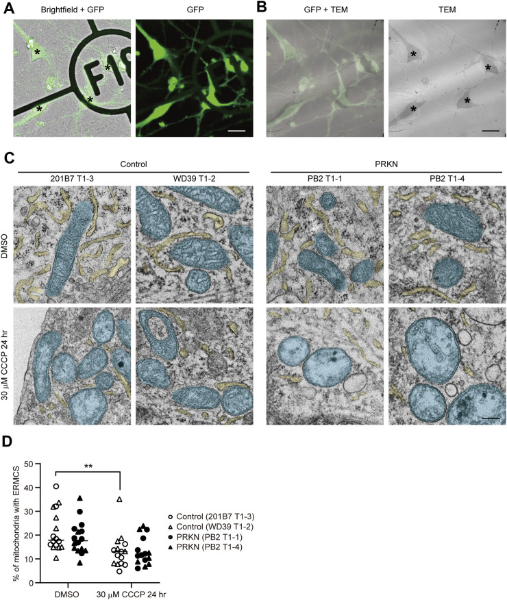
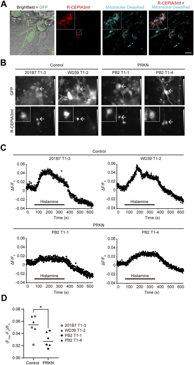
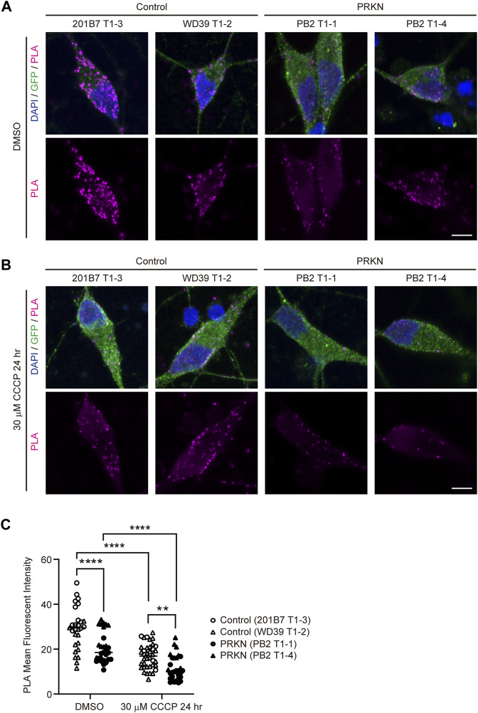
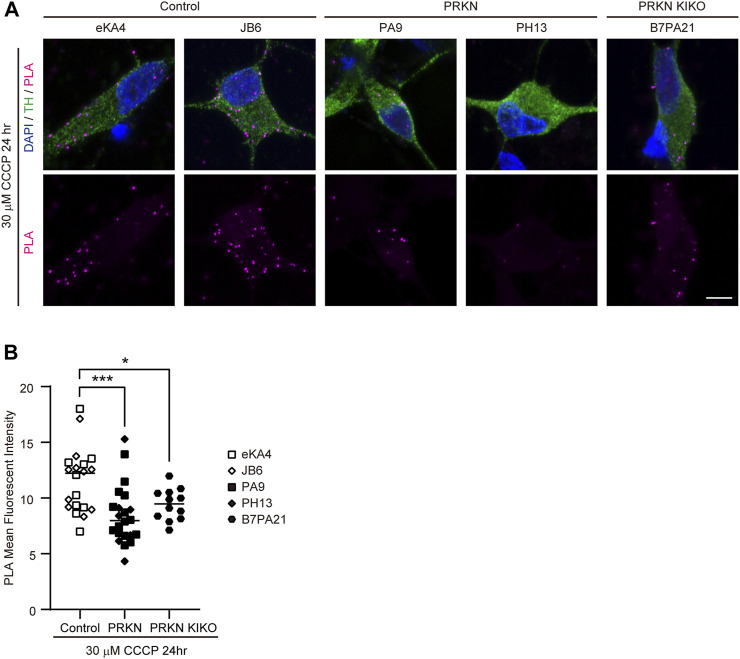
Endoplasmic reticulum-mitochondrial contact sites (ERMCS) play an important role in mitochondrial dynamics, calcium signaling, and autophagy. Disruption of the ERMCS has been linked to several neurodegenerative diseases, including Alzheimer's disease (AD), Parkinson's disease (PD), and amyotrophic lateral sclerosis (ALS). However, the etiological role of ERMCS in these diseases remains unclear. We previously established tyrosine hydroxylase reporter (GFP) iPSC lines from a PD patient with a mutation to perform correlative light-electron microscopy (CLEM) analysis and live cell imaging in GFP-expressing dopaminergic neurons. Here, we analyzed ERMCS in GFP-expressing mutant dopaminergic neurons from patients using CLEM and a proximity ligation assay (PLA). The PLA showed that the ERMCS were significantly reduced in mutant patient dopaminergic neurons compared to the control under normal conditions. The reduction of the ERMCS in -mutant patient dopaminergic neurons was further enhanced by treatment with a mitochondrial uncoupler. In addition, mitochondrial calcium imaging showed that mitochondrial Ca flux was significantly reduced in -mutant patient dopaminergic neurons compared to the control. These results suggest a defect in calcium flux from ER to mitochondria is due to the decreased ERMCS in -mutant patient dopaminergic neurons. Our study of ERMCS using GFP iPSC lines would contribute to further understanding of the mechanisms of dopaminergic neuron degeneration in patients with mutations.
内质网-线粒体接触位点(ERMCS)在线粒体动力学、钙信号传导和自噬中发挥重要作用。ERMCS的破坏与包括阿尔茨海默病(AD)、帕金森病(PD)和肌萎缩侧索硬化症(ALS)在内的几种神经退行性疾病有关。然而,ERMCS在这些疾病中的病因学作用仍不清楚。我们之前从一名患有突变的帕金森病患者建立了酪氨酸羟化酶报告基因(GFP)诱导多能干细胞系,以在表达GFP的多巴胺能神经元中进行相关光电子显微镜(CLEM)分析和活细胞成像。在这里,我们使用CLEM和邻近连接分析(PLA)分析了患者来源的表达GFP的突变多巴胺能神经元中的ERMCS。PLA显示,在正常条件下,与对照组相比,突变患者多巴胺能神经元中的ERMCS显著减少。用线粒体解偶联剂处理后,突变患者多巴胺能神经元中ERMCS的减少进一步加剧。此外,线粒体钙成像显示,与对照组相比,突变患者多巴胺能神经元中的线粒体钙通量显著降低。这些结果表明,突变患者多巴胺能神经元中从内质网到线粒体的钙通量缺陷是由于ERMCS减少所致。我们使用GFP诱导多能干细胞系对ERMCS的研究将有助于进一步了解携带突变的患者中多巴胺能神经元变性的机制。