Department of Radiology, Pitié-Salpêtrière Hospital, Assistance Publique-Hôpitaux de Paris (AP-HP), Paris, France.
Clinical Research Unit, Pitié-Salpêtrière Hospital, Assistance Publique-Hôpitaux de Paris (AP-HP), Paris, France.
Cancer Med. 2023 Oct;12(19):19500-19511. doi: 10.1002/cam4.6560. Epub 2023 Sep 29.
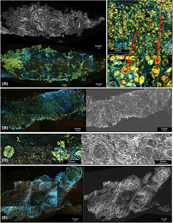
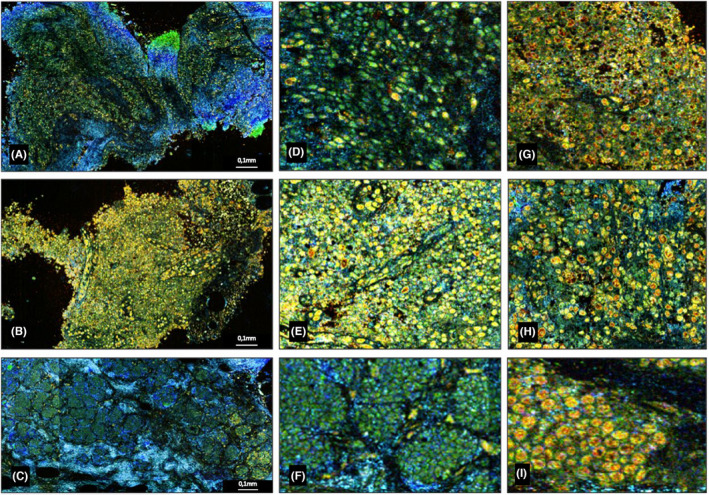
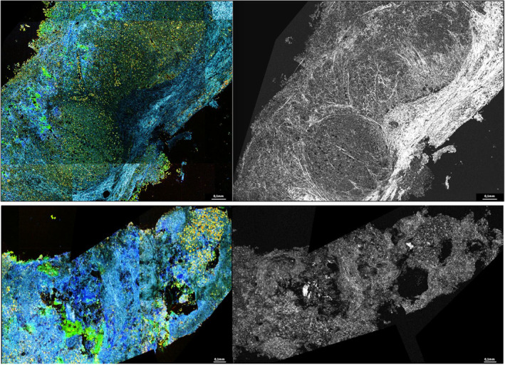
Full-field optical coherence tomography combined with dynamic cell imaging (D-FFOCT) is a new, simple-to-use, nondestructive, quick technique that can provide sufficient spatial resolution to mimic histopathological analysis. The objective of this study was to evaluate diagnostic performance of D-FFOCT for one-stop rapid diagnosis breast clinic.
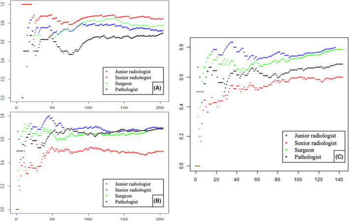
Dynamic full-field optical coherence tomography was applied to fresh, untreated breast and nodes biopsies. Four different readers (senior and junior radiologist, surgeon, and pathologist) analyzed the samples without knowing final histological diagnosis or American College of Radiology classification. The results were compared to conventional processing and staining (hematoxylin-eosin).
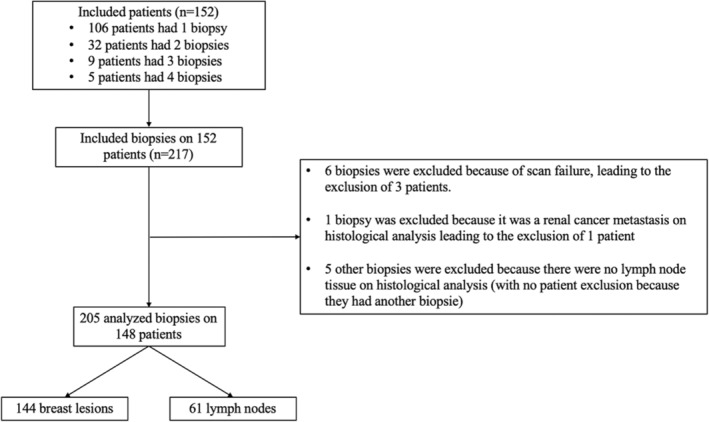
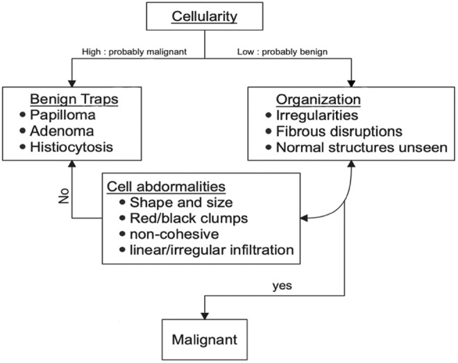
A total of 217 biopsies were performed on 152 patients. There were 144 breast biopsies and 61 lymph nodes with 101 infiltrative cancers (49.27%), 99 benign lesions (48.29%), 3 ductal in situ carcinoma (1.46%), and 2 atypias (0.98%). The diagnostic performance results were as follow: sensitivity: 77% [0.7;0.82], specificity: 64% [0.58;0.71], PPV: 74% [0.68;0.78], and NPV: 75% [0.72;0.78]. A large image atlas was created as well as a diagnosis algorithm from the readers' experience.
With 74% PPV and 75% NPV, D-FFOCT is not yet ready to be used in clinical practice to identify breast cancer. This is mainly explained by the lack of experience and knowledge of this new technic by the four lectors. By training with the diagnosis algorithm and the image atlas, radiologists could have better outcomes allowing quick detection of breast cancer and lymph node involvement. Deep learning could also be used, and further investigation will follow.
全场光学相干断层成像术结合动态细胞成像(D-FFOCT)是一种新的、易于使用的、非破坏性的、快速的技术,它可以提供足够的空间分辨率来模拟组织病理学分析。本研究的目的是评估 D-FFOCT 在一站式快速诊断乳房诊所中的诊断性能。
动态全场光学相干断层成像术应用于新鲜、未经处理的乳房和淋巴结活检。四位不同的读者(资深和初级放射科医生、外科医生和病理科医生)在不知道最终组织学诊断或美国放射学院分类的情况下分析样本。结果与常规处理和染色(苏木精-伊红)进行比较。
共对 152 名患者的 217 个活检进行了检查。其中有 144 个乳房活检和 61 个淋巴结活检,包括 101 例浸润性癌(49.27%)、99 例良性病变(48.29%)、3 例导管原位癌(1.46%)和 2 例不典型病变(0.98%)。诊断性能结果如下:敏感性:77%[0.7;0.82],特异性:64%[0.58;0.71],PPV:74%[0.68;0.78],NPV:75%[0.72;0.78]。创建了一个大型图像图谱以及根据读者经验的诊断算法。
D-FFOCT 的 PPV 为 74%,NPV 为 75%,尚未准备好用于临床实践以识别乳腺癌。这主要是由于四位读者缺乏对这种新技术的经验和知识。通过使用诊断算法和图像图谱进行培训,放射科医生可以更好地进行检测,从而快速发现乳腺癌和淋巴结受累。还可以使用深度学习,进一步的研究将随之进行。