Bianconi Andrea, Rossi Luca Francesco, Bonada Marta, Zeppa Pietro, Nico Elsa, De Marco Raffaele, Lacroce Paola, Cofano Fabio, Bruno Francesco, Morana Giovanni, Melcarne Antonio, Ruda Roberta, Mainardi Luca, Fiaschi Pietro, Garbossa Diego, Morra Lia
Neurosurgery, Department of Neuroscience, University of Turin, via Cherasco 15, 10126, Turin, Italy.
Dipartimento di Automatica e Informatica, Politecnico di Torino, Turin, Italy.
Brain Inform. 2023 Oct 6;10(1):26. doi: 10.1186/s40708-023-00207-6.
Clinical and surgical decisions for glioblastoma patients depend on a tumor imaging-based evaluation. Artificial Intelligence (AI) can be applied to magnetic resonance imaging (MRI) assessment to support clinical practice, surgery planning and prognostic predictions. In a real-world context, the current obstacles for AI are low-quality imaging and postoperative reliability. The aim of this study is to train an automatic algorithm for glioblastoma segmentation on a clinical MRI dataset and to obtain reliable results both pre- and post-operatively.
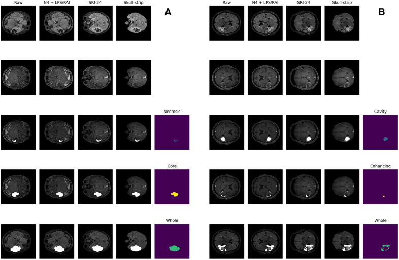
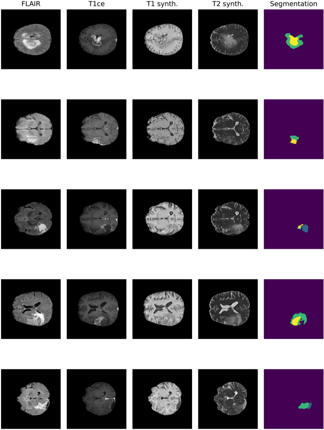
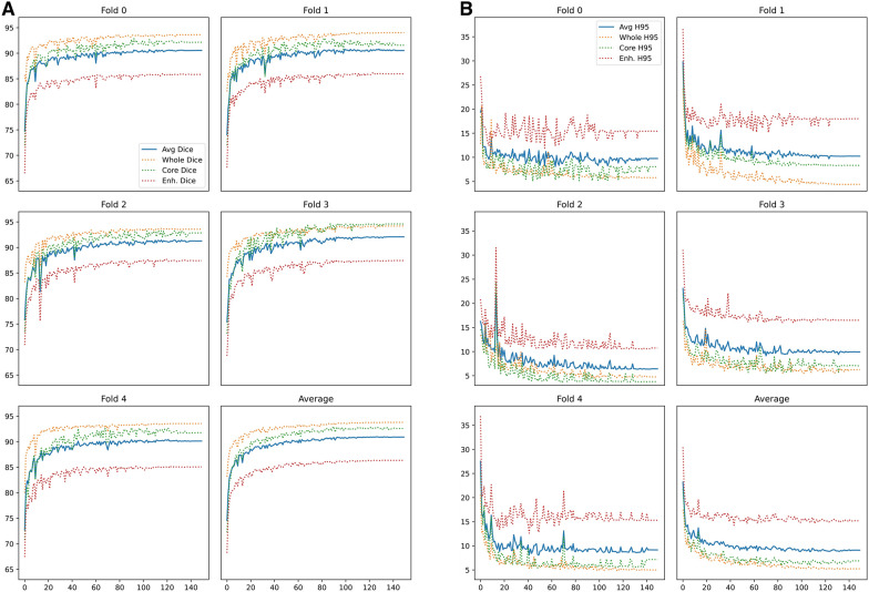
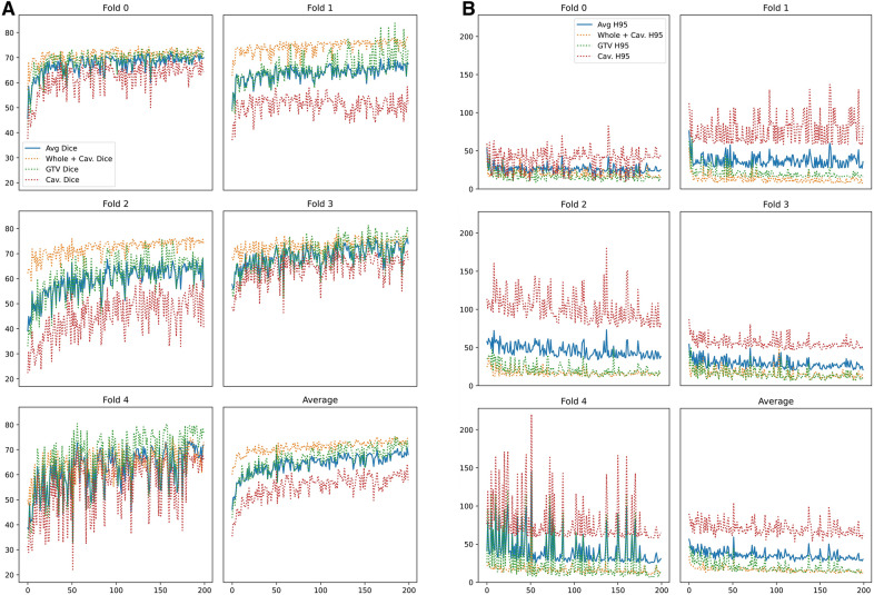
The dataset used for this study comprises 237 (71 preoperative and 166 postoperative) MRIs from 71 patients affected by a histologically confirmed Grade IV Glioma. The implemented U-Net architecture was trained by transfer learning to perform the segmentation task on postoperative MRIs. The training was carried out first on BraTS2021 dataset for preoperative segmentation. Performance is evaluated using DICE score (DS) and Hausdorff 95% (H95).
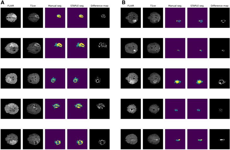
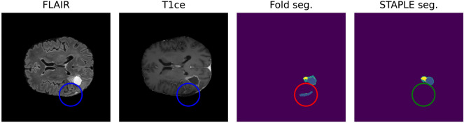
In preoperative scenario, overall DS is 91.09 (± 0.60) and H95 is 8.35 (± 1.12), considering tumor core, enhancing tumor and whole tumor (ET and edema). In postoperative context, overall DS is 72.31 (± 2.88) and H95 is 23.43 (± 7.24), considering resection cavity (RC), gross tumor volume (GTV) and whole tumor (WT). Remarkably, the RC segmentation obtained a mean DS of 63.52 (± 8.90) in postoperative MRIs.
The performances achieved by the algorithm are consistent with previous literature for both pre-operative and post-operative glioblastoma's MRI evaluation. Through the proposed algorithm, it is possible to reduce the impact of low-quality images and missing sequences.
胶质母细胞瘤患者的临床和手术决策取决于基于肿瘤成像的评估。人工智能(AI)可应用于磁共振成像(MRI)评估,以支持临床实践、手术规划和预后预测。在实际应用中,AI目前面临的障碍是成像质量低和术后可靠性问题。本研究的目的是在临床MRI数据集上训练一种用于胶质母细胞瘤分割的自动算法,并在术前和术后均获得可靠结果。
本研究使用的数据集包括来自71例经组织学确诊为IV级胶质瘤患者的237张MRI图像(71例术前和166例术后)。通过迁移学习训练所实现的U-Net架构,以在术后MRI图像上执行分割任务。首先在BraTS2021数据集上进行术前分割训练。使用DICE评分(DS)和Hausdorff 95%(H95)评估性能。
在术前情况下,考虑肿瘤核心、强化肿瘤和整个肿瘤(增强肿瘤和水肿),总体DS为91.09(±0.60),H95为8.35(±1.12)。在术后情况下,考虑切除腔(RC)、大体肿瘤体积(GTV)和整个肿瘤(WT),总体DS为72.31(±2.88),H95为23.43(±7.24)。值得注意的是,术后MRI图像中RC分割的平均DS为63.52(±8.90)。
该算法在术前和术后胶质母细胞瘤的MRI评估中所取得的性能与先前文献一致。通过所提出的算法,可以减少低质量图像和缺失序列的影响。