Department of Dermatology, University of California, Davis, CA, 95616, USA.
Department of Ophthalmology, University of California, Davis, CA, 95616, USA.
Sci Rep. 2023 Oct 6;13(1):16885. doi: 10.1038/s41598-023-42743-5.
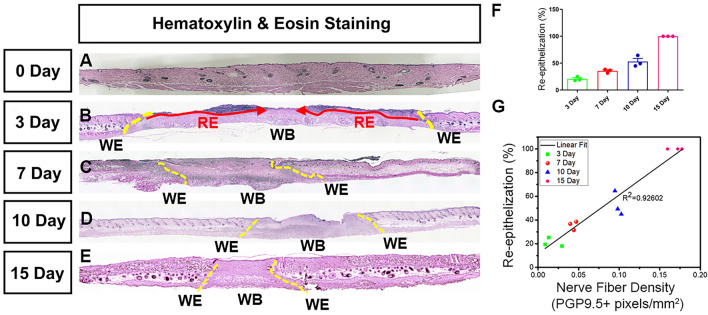
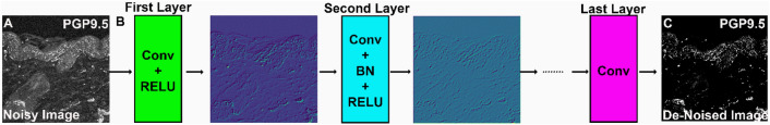
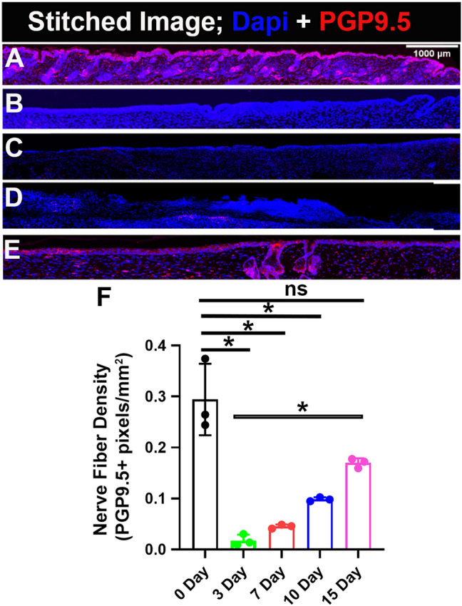
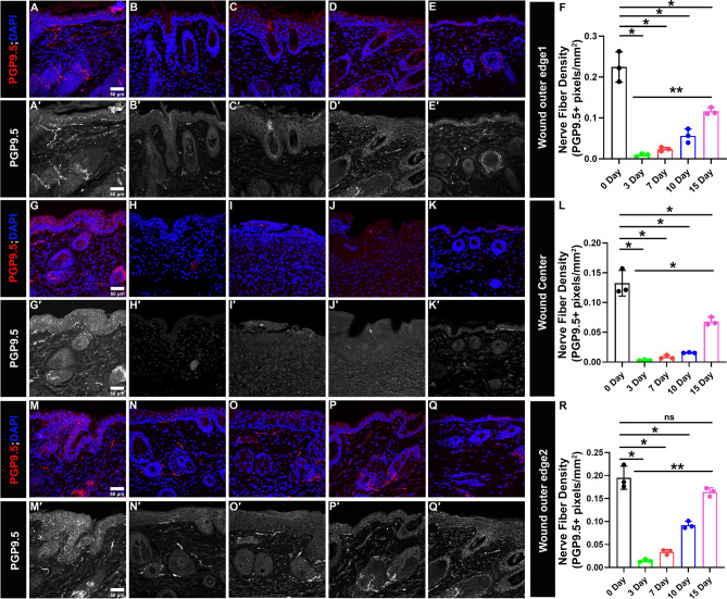
The peripheral nerves (PNs) innervate the dermis and epidermis, and are suggested to play an important role in wound healing. Several methods to quantify skin innervation during wound healing have been reported. Those usually require multiple observers, are complex and labor-intensive, and the noise/background associated with the immunohistochemistry (IHC) images could cause quantification errors/user bias. In this study, we employed the state-of-the-art deep neural network, Denoising Convolutional Neural Network (DnCNN), to perform pre-processing and effectively reduce the noise in the IHC images. Additionally, we utilized an automated image analysis tool, assisted by Matlab, to accurately determine the extent of skin innervation during various stages of wound healing. The 8 mm wound is generated using a circular biopsy punch in the wild-type mouse. Skin samples were collected on days 3, 7, 10 and 15, and sections from paraffin-embedded tissues were stained against pan-neuronal marker- protein-gene-product 9.5 (PGP 9.5) antibody. On day 3 and day 7, negligible nerve fibers were present throughout the wound with few only on the lateral boundaries of the wound. On day 10, a slight increase in nerve fiber density appeared, which significantly increased on day 15. Importantly, we found a positive correlation (R = 0.926) between nerve fiber density and re-epithelization, suggesting an association between re-innervation and re-epithelization. These results established a quantitative time course of re-innervation in wound healing, and the automated image analysis method offers a novel and useful tool to facilitate the quantification of innervation in the skin and other tissues.
周围神经(PNs)支配真皮和表皮,并被认为在伤口愈合中发挥重要作用。已经报道了几种量化伤口愈合过程中皮肤神经支配的方法。这些方法通常需要多个观察者,且复杂且劳动强度大,免疫组织化学(IHC)图像相关的噪声/背景可能会导致定量错误/用户偏见。在这项研究中,我们采用了最先进的深度神经网络,去噪卷积神经网络(DnCNN),对 IHC 图像进行预处理,有效地降低了噪声。此外,我们利用了一个自动化的图像分析工具,由 Matlab 辅助,在伤口愈合的各个阶段准确地确定皮肤神经支配的程度。在野生型小鼠中,使用圆形活检冲孔产生 8mm 伤口。在第 3、7、10 和 15 天收集皮肤样本,并对石蜡包埋组织切片进行针对泛神经元标志物-蛋白基因产物 9.5(PGP 9.5)抗体的染色。在第 3 天和第 7 天,整个伤口中几乎没有神经纤维,只有少数位于伤口的外侧边界。在第 10 天,神经纤维密度略有增加,第 15 天明显增加。重要的是,我们发现神经纤维密度与再上皮化之间存在正相关(R=0.926),这表明再神经支配与再上皮化之间存在关联。这些结果确立了伤口愈合中再神经支配的定量时间过程,并且自动化的图像分析方法为量化皮肤和其他组织中的神经支配提供了一种新颖而有用的工具。