Department of Chemistry and Bio-X, Stanford University, Stanford, CA, USA.
School of Medicine, Stanford University, Stanford, CA, USA.
Nat Nanotechnol. 2022 Jun;17(6):653-660. doi: 10.1038/s41565-022-01130-3. Epub 2022 May 23.
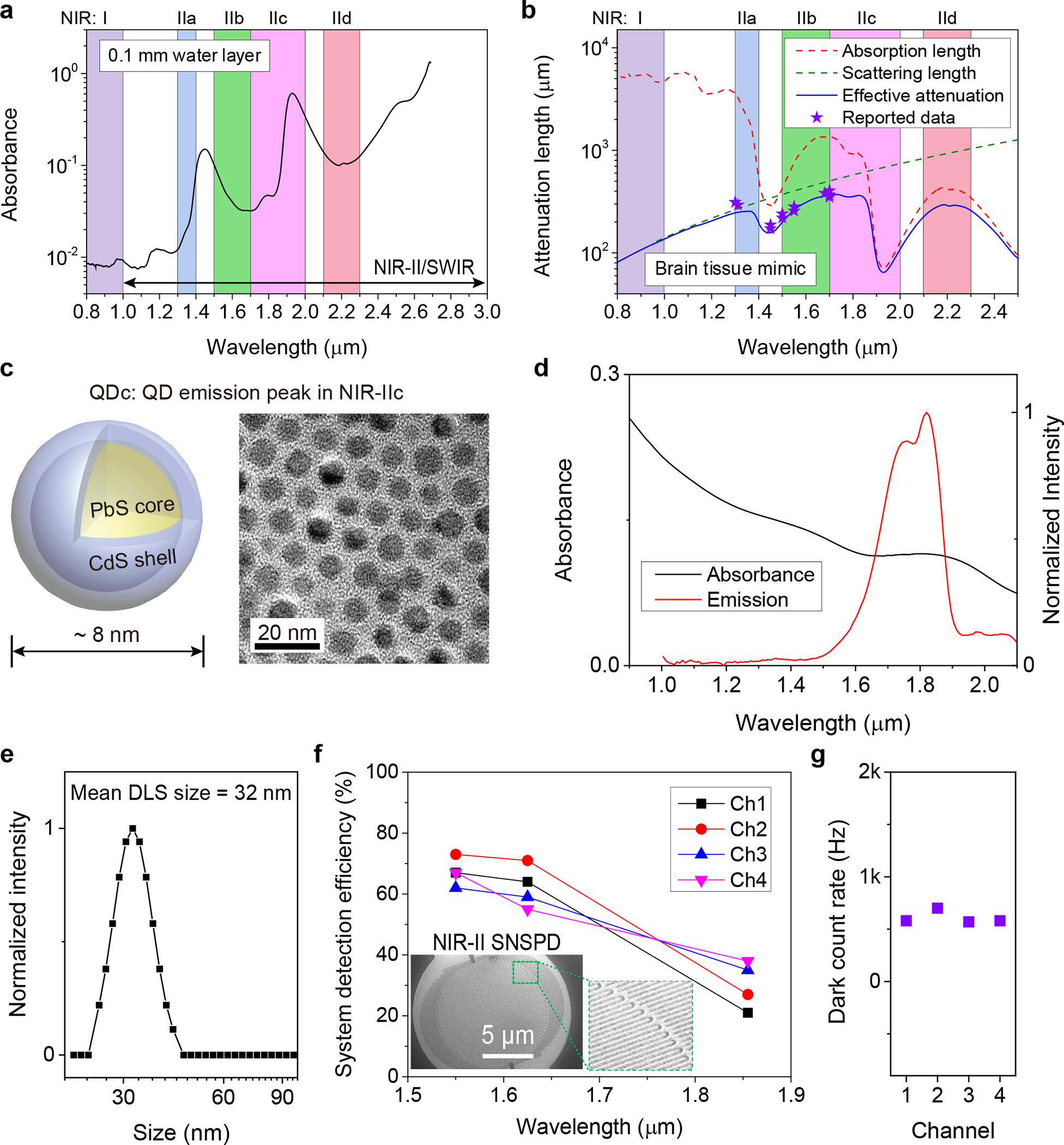
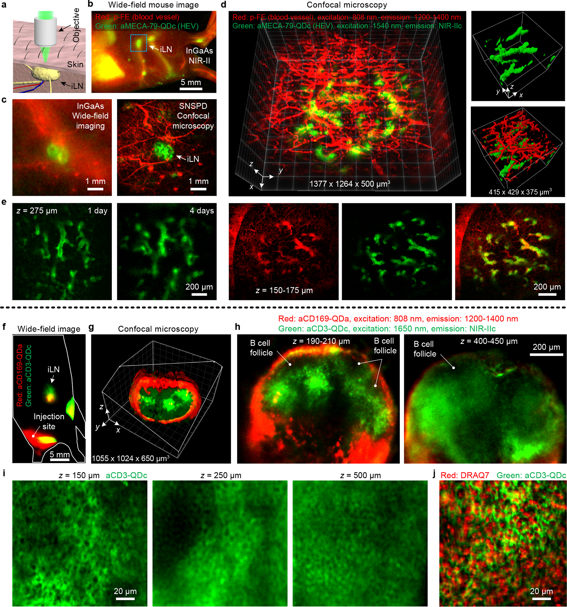
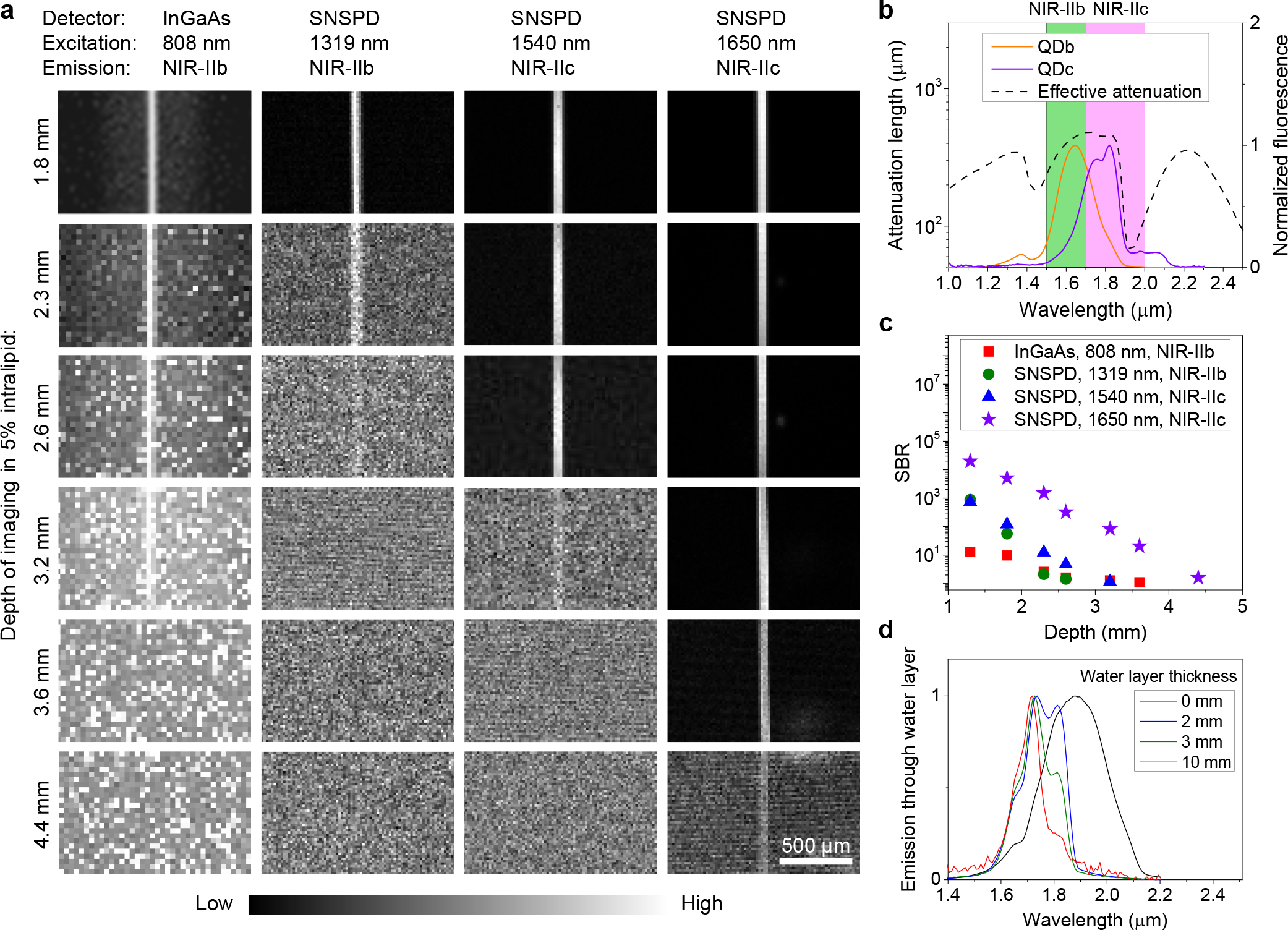
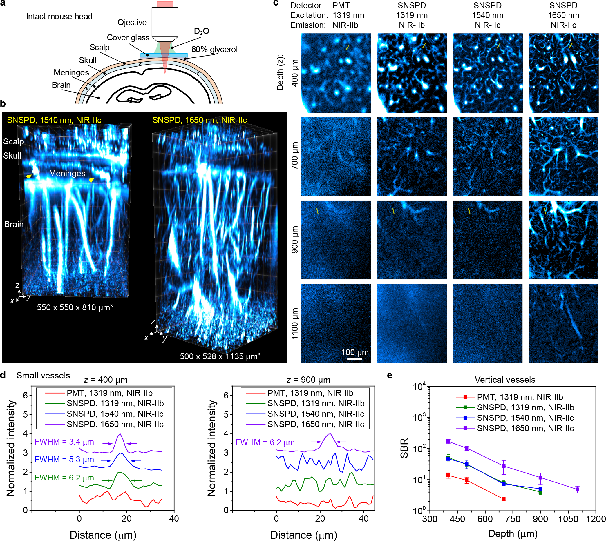
Light scattering by biological tissues sets a limit to the penetration depth of high-resolution optical microscopy imaging of live mammals in vivo. An effective approach to reduce light scattering and increase imaging depth is to extend the excitation and emission wavelengths to the second near-infrared window (NIR-II) at >1,000 nm, also called the short-wavelength infrared window. Here we show biocompatible core-shell lead sulfide/cadmium sulfide quantum dots emitting at ~1,880 nm and superconducting nanowire single-photon detectors for single-photon detection up to 2,000 nm, enabling a one-photon excitation fluorescence imaging window in the 1,700-2,000 nm (NIR-IIc) range with 1,650 nm excitation-the longest one-photon excitation and emission for in vivo mouse imaging so far. Confocal fluorescence imaging in NIR-IIc reached an imaging depth of ~1,100 μm through an intact mouse head, and enabled non-invasive cellular-resolution imaging in the inguinal lymph nodes of mice without any surgery. We achieve in vivo molecular imaging of high endothelial venules with diameters as small as ~6.6 μm, as well as CD169 + macrophages and CD3 + T cells in the lymph nodes, opening the possibility of non-invasive intravital imaging of immune trafficking in lymph nodes at the single-cell/vessel-level longitudinally.
生物组织的光散射限制了活体哺乳动物高分辨率光学显微镜成像的穿透深度。减少光散射并增加成像深度的一种有效方法是将激发和发射波长扩展到>1000nm 的第二个近红外窗口(NIR-II),也称为短波红外窗口。在这里,我们展示了发射波长约为 1880nm 的生物相容性核壳型硫化铅/硫化镉量子点和超导纳米线单光子探测器,用于单光子探测至 2000nm,从而在 1700-2000nm(NIR-IIc)范围内实现了单光子激发荧光成像窗口,激发和发射波长最长为 1650nm-这是迄今为止活体小鼠成像的最长单光子激发和发射。通过完整的小鼠头部,在 NIR-IIc 中进行共聚焦荧光成像,实现了1100μm 的成像深度,并且可以在不进行任何手术的情况下对小鼠腹股沟淋巴结进行非侵入性的细胞分辨率成像。我们实现了高内皮小静脉(直径小至6.6μm)以及淋巴结中 CD169+巨噬细胞和 CD3+T 细胞的体内分子成像,为单细胞/血管水平的淋巴结内免疫运输的非侵入性活体成像提供了可能。