Zhang Le, Antonacci Michael, Burant Alex, McCallister Andrew, Kelley Michele, Bryden Nicholas, McHugh Christian, Atalla Sebastian, Holmes Leah, Katz Laurence, Branca Rosa Tamara
Department of Physics and Astronomy, University of North Carolina at Chapel Hill, 27599, Chapel Hill, NC, USA.
Biomedical Research Imaging Center, University of North Carolina at Chapel Hill, 27599, Chapel Hill, NC, USA.
Commun Med (Lond). 2023 Oct 17;3(1):147. doi: 10.1038/s43856-023-00374-x.
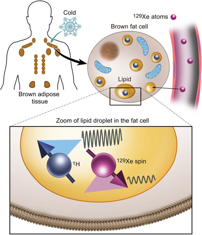
Absolute temperature measurements of tissues inside the human body are difficult to perform non-invasively. Yet, for brown adipose tissue (BAT), these measurements would enable direct monitoring of its thermogenic activity and its association with metabolic health.
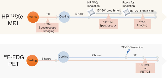
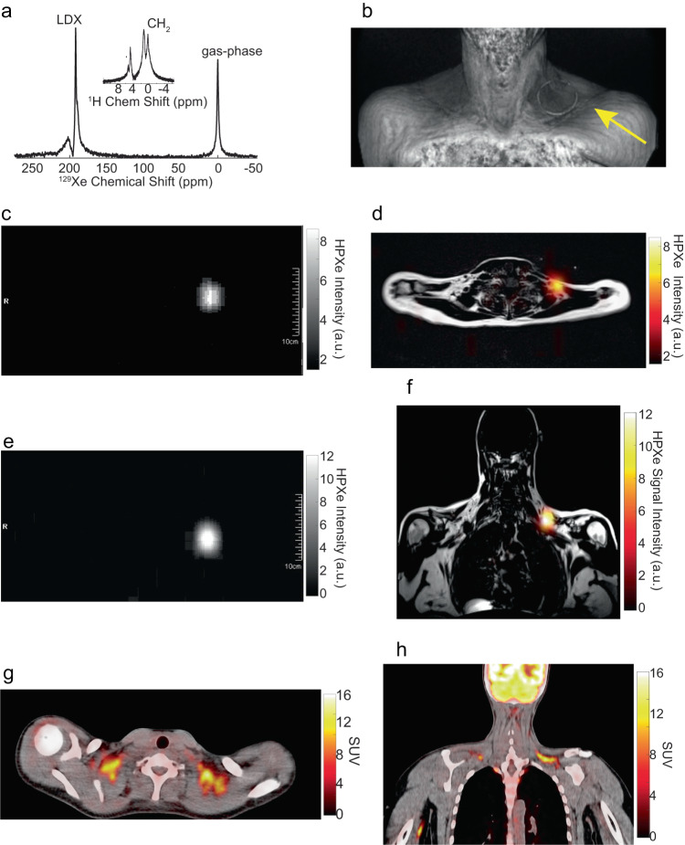
Here, we report direct measurement of absolute BAT temperature in humans during cold exposure by magnetic resonance (MR) with laser polarized xenon gas. This methodology, which leverages on the sensitivity of the chemical shift of the Xe isotope to temperature-induced changes in fat density, is first calibrated in vitro and then tested in vivo in rodents. Finally, it is used in humans along with positron emission tomography (PET) scans with fluorine-18-fluorodeoxyglucose to detect BAT thermogenic activity during cold exposure.
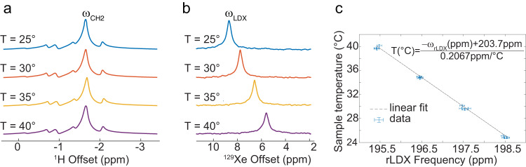
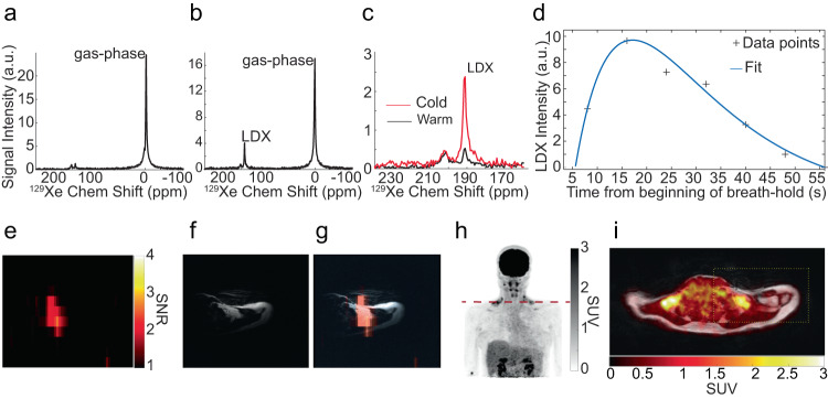
Absolute temperature measurements, obtained in rodents with an experimental error of 0.5 °C, show only a median deviation of 0.12 °C against temperature measurements made using a pre-calibrated optical temperature probe. In humans, enhanced uptake of Xe in BAT during cold exposure leads to background-free detection of this tissue by MR. Global measurements of supraclavicular BAT temperature, made over the course of four seconds and with an experimental error ranging from a minimum of 0.4 °C to more than 2 °C, in case of poor shimming, reveal an average BAT temperature of 38.8° ± 0.8 °C, significantly higher (p < 0.02 two-sided t test) than 37.7 °C. Hot BAT is also detected in participants with a PET scan negative for BAT.
Non-invasive, radiation-free measurements of BAT temperature by MRI with hyperpolarized Xe may enable longitudinal monitoring of human BAT activity under various stimulatory conditions.
对人体内部组织进行绝对温度测量很难通过非侵入性方式实现。然而,对于棕色脂肪组织(BAT)而言,这些测量能够直接监测其产热活性及其与代谢健康的关联。
在此,我们报告了通过激光极化氙气磁共振(MR)对人体在冷暴露期间的BAT绝对温度进行直接测量。这种利用氙同位素化学位移对温度引起的脂肪密度变化的敏感性的方法,首先在体外进行校准,然后在啮齿动物体内进行测试。最后,将其与使用氟 - 18 - 氟脱氧葡萄糖的正电子发射断层扫描(PET)一起用于人体,以检测冷暴露期间的BAT产热活性。
在啮齿动物中获得的绝对温度测量结果,实验误差为0.5°C,与使用预先校准的光学温度探头进行的温度测量相比,中位偏差仅为0.12°C。在人体中,冷暴露期间BAT中氙的摄取增加,使得通过MR能够无背景地检测到该组织。在四秒内进行的锁骨上BAT温度的整体测量,实验误差范围从最小0.4°C到超过2°C(在匀场不佳的情况下),显示BAT平均温度为38.8°±0.8°C,显著高于37.7°C(双侧t检验p < 0.02)。在PET扫描显示BAT阴性的参与者中也检测到了热BAT。
通过超极化氙气MRI对BAT温度进行无创、无辐射测量,可能有助于在各种刺激条件下对人体BAT活动进行纵向监测。