Feng Ruibin, Deb Brototo, Ganesan Prasanth, Tjong Fleur V Y, Rogers Albert J, Ruipérez-Campillo Samuel, Somani Sulaiman, Clopton Paul, Baykaner Tina, Rodrigo Miguel, Zou James, Haddad Francois, Zahari Matei, Narayan Sanjiv M
Department of Medicine and Cardiovascular Institute, Stanford University, Stanford, CA, United States.
Heart Center, Department of Clinical and Experimental Cardiology, Amsterdam UMC, University of Amsterdam, Amsterdam, Netherlands.
Front Cardiovasc Med. 2023 Oct 2;10:1189293. doi: 10.3389/fcvm.2023.1189293. eCollection 2023.
Segmentation of computed tomography (CT) is important for many clinical procedures including personalized cardiac ablation for the management of cardiac arrhythmias. While segmentation can be automated by machine learning (ML), it is limited by the need for large, labeled training data that may be difficult to obtain. We set out to combine ML of cardiac CT with domain knowledge, which reduces the need for large training datasets by encoding cardiac geometry, which we then tested in independent datasets and in a prospective study of atrial fibrillation (AF) ablation.
We mathematically represented atrial anatomy with simple geometric shapes and derived a model to parse cardiac structures in a small set of = 6 digital hearts. The model, termed "virtual dissection," was used to train ML to segment cardiac CT in = 20 patients, then tested in independent datasets and in a prospective study.
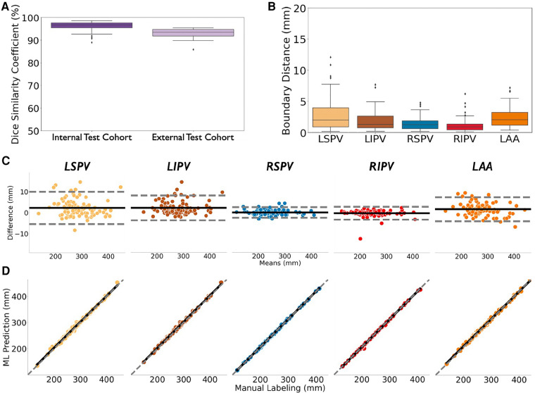
In independent test cohorts ( = 160) from 2 Institutions with different CT scanners, atrial structures were accurately segmented with Dice scores of 96.7% in internal (IQR: 95.3%-97.7%) and 93.5% in external (IQR: 91.9%-94.7%) test data, with good agreement with experts ( = 0.99; < 0.0001). In a prospective study of 42 patients at ablation, this approach reduced segmentation time by 85% (2.3 ± 0.8 vs. 15.0 ± 6.9 min, < 0.0001), yet provided similar Dice scores to experts (93.9% (IQR: 93.0%-94.6%) vs. 94.4% (IQR: 92.8%-95.7%), = NS).
Encoding cardiac geometry using mathematical models greatly accelerated training of ML to segment CT, reducing the need for large training sets while retaining accuracy in independent test data. Combining ML with domain knowledge may have broad applications.
计算机断层扫描(CT)分割对于许多临床程序都很重要,包括用于心律失常管理的个性化心脏消融。虽然分割可以通过机器学习(ML)实现自动化,但它受到对可能难以获得的大量标记训练数据的需求的限制。我们着手将心脏CT的机器学习与领域知识相结合,通过对心脏几何结构进行编码来减少对大型训练数据集的需求,然后我们在独立数据集和心房颤动(AF)消融的前瞻性研究中对其进行了测试。
我们用简单的几何形状对心房解剖结构进行数学表示,并推导了一个模型来解析一小组(n = 6)数字心脏中的心脏结构。该模型被称为“虚拟解剖”,用于训练机器学习以分割20例患者的心脏CT,然后在独立数据集和前瞻性研究中进行测试。
在来自2个使用不同CT扫描仪的机构的独立测试队列(n = 160)中,内部测试数据(IQR:95.3% - 97.7%)的心房结构分割准确率为96.7%,外部测试数据(IQR:91.9% - 94.7%)的准确率为93.5%,与专家的一致性良好(κ = 0.99;P < 0.0001)。在对42例进行消融治疗的患者的前瞻性研究中,这种方法将分割时间减少了85%(2.3 ± 0.8分钟对15.0 ± 6.9分钟,P < 0.0001),但与专家的Dice评分相似(93.9%(IQR:93.0% - 94.6%)对94.4%(IQR:92.8% - 95.7%),P = NS)。
使用数学模型对心脏几何结构进行编码极大地加速了用于分割CT的机器学习的训练,减少了对大型训练集的需求,同时在独立测试数据中保持了准确性。将机器学习与领域知识相结合可能具有广泛的应用。