• 文献检索
  • 文档翻译
  • 深度研究
  • 学术资讯
  • Suppr Zotero 插件Zotero 插件
  • 邀请有礼
  • 套餐&价格
  • 历史记录
应用&插件
Suppr Zotero 插件Zotero 插件浏览器插件Mac 客户端Windows 客户端微信小程序
定价
高级版会员购买积分包购买API积分包
服务
文献检索文档翻译深度研究API 文档MCP 服务
关于我们
关于 Suppr公司介绍联系我们用户协议隐私条款
关注我们

Suppr 超能文献

核心技术专利:CN118964589B侵权必究
粤ICP备2023148730 号-1Suppr @ 2026

文献检索

告别复杂PubMed语法,用中文像聊天一样搜索,搜遍4000万医学文献。AI智能推荐,让科研检索更轻松。

立即免费搜索

文件翻译

保留排版,准确专业,支持PDF/Word/PPT等文件格式,支持 12+语言互译。

免费翻译文档

深度研究

AI帮你快速写综述,25分钟生成高质量综述,智能提取关键信息,辅助科研写作。

立即免费体验

深紫外显微镜鉴定前列腺基底细胞:前列腺癌诊断的重要生物标志物

Deep UV Microscopy Identifies Prostatic Basal Cells: An Important Biomarker for Prostate Cancer Diagnostics.

作者信息

Soltani Soheil, Cheng Brian, Osunkoya Adeboye O, Robles Francisco E

机构信息

Wallace H. Coulter Department of Biomedical Engineering, Georgia Institute of Technology and Emory University, Atlanta, GA 30332, USA.

Departments of Pathology and Urology, Emory University School of Medicine, Atlanta, GA 30322, USA.

出版信息

BME Front. 2022 Sep 2;2022:9847962. doi: 10.34133/2022/9847962. eCollection 2022.

DOI:10.34133/2022/9847962
PMID:37850167
原文链接:https://pmc.ncbi.nlm.nih.gov/articles/PMC10521648/
Abstract

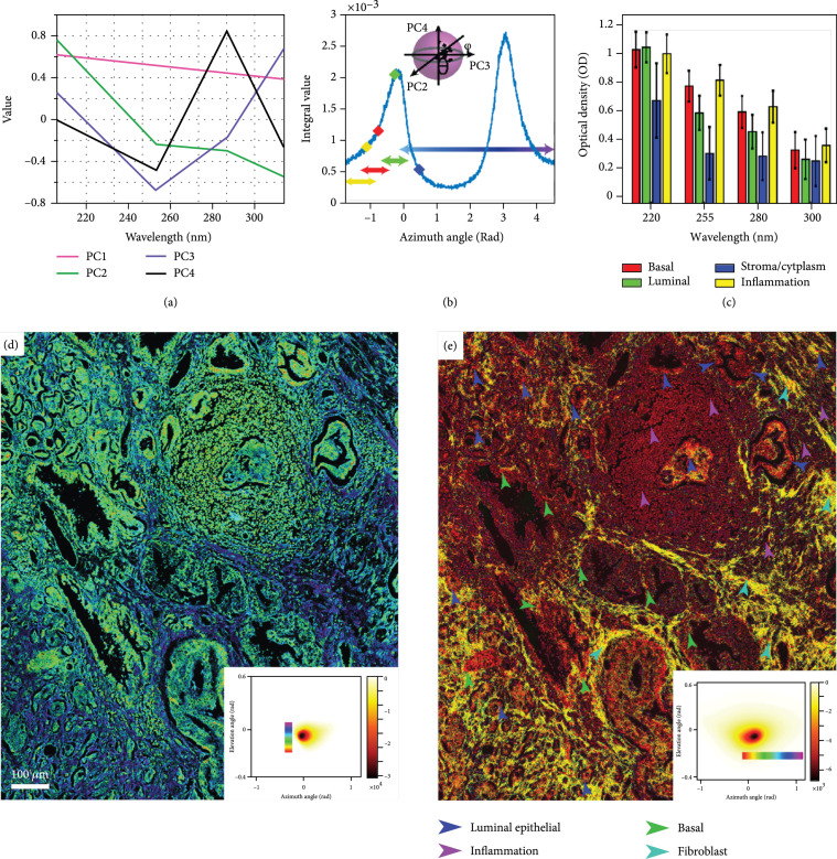
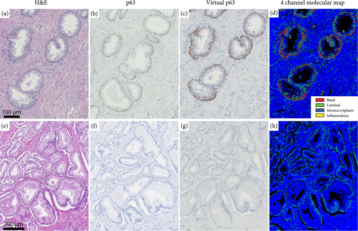
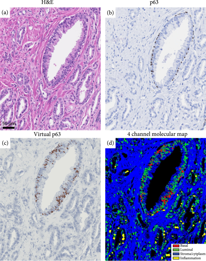
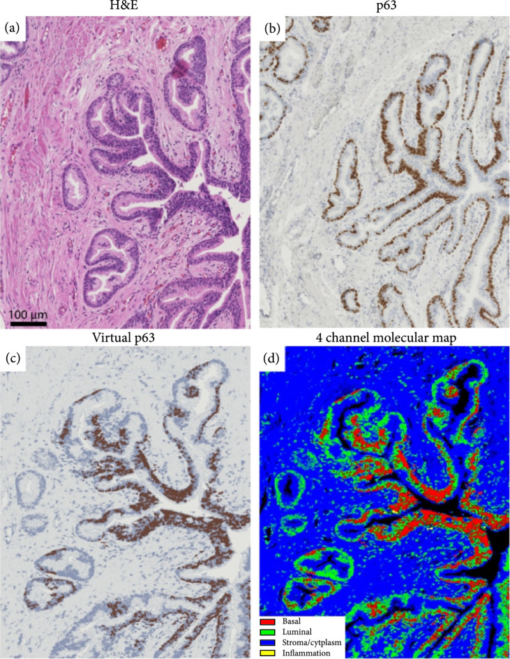
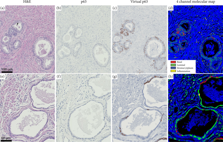
. Identifying benign mimics of prostatic adenocarcinoma remains a significant diagnostic challenge. In this work, we developed an approach based on label-free, high-resolution molecular imaging with multispectral deep ultraviolet (UV) microscopy which identifies important prostate tissue components, including basal cells. This work has significant implications towards improving the pathologic assessment and diagnosis of prostate cancer. . One of the most important indicators of prostate cancer is the absence of basal cells in glands and ducts. However, identifying basal cells using hematoxylin and eosin (H&E) stains, which is the standard of care, can be difficult in a subset of cases. In such situations, pathologists often resort to immunohistochemical (IHC) stains for a definitive diagnosis. However, IHC is expensive and time-consuming and requires more tissue sections which may not be available. In addition, IHC is subject to false-negative or false-positive stains which can potentially lead to an incorrect diagnosis. . We leverage the rich molecular information of label-free multispectral deep UV microscopy to uniquely identify basal cells, luminal cells, and inflammatory cells. The method applies an unsupervised geometrical representation of principal component analysis to separate the various components of prostate tissue leading to multiple image representations of the molecular information. . Our results show that this method accurately and efficiently identifies benign and malignant glands with high fidelity, free of any staining procedures, based on the presence or absence of basal cells. We further use the molecular information to directly generate a high-resolution virtual IHC stain that clearly identifies basal cells, even in cases where IHC stains fail. . Our simple, low-cost, and label-free deep UV method has the potential to improve and facilitate prostate cancer diagnosis by enabling robust identification of basal cells and other important prostate tissue components.

摘要

识别前列腺腺癌的良性模拟物仍然是一项重大的诊断挑战。在这项工作中,我们开发了一种基于无标记、高分辨率分子成像的方法,采用多光谱深紫外(UV)显微镜来识别重要的前列腺组织成分,包括基底细胞。这项工作对改善前列腺癌的病理评估和诊断具有重要意义。前列腺癌最重要的指标之一是腺体和导管中不存在基底细胞。然而,使用苏木精和伊红(H&E)染色(这是标准的诊断方法)来识别基底细胞,在某些情况下可能会很困难。在这种情况下,病理学家通常会采用免疫组织化学(IHC)染色来进行明确诊断。然而,IHC成本高、耗时,并且需要更多可能无法获得的组织切片。此外,IHC可能会出现假阴性或假阳性染色,这可能会导致错误的诊断。我们利用无标记多光谱深紫外显微镜丰富的分子信息来独特地识别基底细胞、管腔细胞和炎症细胞。该方法应用主成分分析的无监督几何表示来分离前列腺组织的各种成分,从而得到分子信息的多种图像表示。我们的结果表明,基于基底细胞的存在与否,该方法能够准确、高效地以高保真度识别良性和恶性腺体,无需任何染色程序。我们进一步利用分子信息直接生成高分辨率的虚拟IHC染色,即使在IHC染色失败的情况下也能清晰地识别基底细胞。我们简单、低成本且无标记的深紫外方法有潜力通过可靠地识别基底细胞和其他重要的前列腺组织成分来改善和促进前列腺癌的诊断。

https://cdn.ncbi.nlm.nih.gov/pmc/blobs/99cd/10521648/163fbd58fc2b/9847962.fig.006.jpg
https://cdn.ncbi.nlm.nih.gov/pmc/blobs/99cd/10521648/6f0b77633259/9847962.fig.001.jpg
https://cdn.ncbi.nlm.nih.gov/pmc/blobs/99cd/10521648/d88c25bce8bc/9847962.fig.002.jpg
https://cdn.ncbi.nlm.nih.gov/pmc/blobs/99cd/10521648/09c59640f07f/9847962.fig.003.jpg
https://cdn.ncbi.nlm.nih.gov/pmc/blobs/99cd/10521648/5366c6194f43/9847962.fig.004.jpg
https://cdn.ncbi.nlm.nih.gov/pmc/blobs/99cd/10521648/3330623b666b/9847962.fig.005.jpg
https://cdn.ncbi.nlm.nih.gov/pmc/blobs/99cd/10521648/163fbd58fc2b/9847962.fig.006.jpg

相似文献

1
Deep UV Microscopy Identifies Prostatic Basal Cells: An Important Biomarker for Prostate Cancer Diagnostics.深紫外显微镜鉴定前列腺基底细胞:前列腺癌诊断的重要生物标志物
BME Front. 2022 Sep 2;2022:9847962. doi: 10.34133/2022/9847962. eCollection 2022.
2
Prostate cancer histopathology using label-free multispectral deep-UV microscopy quantifies phenotypes of tumor aggressiveness and enables multiple diagnostic virtual stains.基于无标记多光谱深紫外显微镜的前列腺癌组织病理学可定量评估肿瘤侵袭性表型,并实现多种诊断性虚拟染色。
Sci Rep. 2022 Jun 4;12(1):9329. doi: 10.1038/s41598-022-13332-9.
3
How often does alpha-methylacyl-CoA-racemase contribute to resolving an atypical diagnosis on prostate needle biopsy beyond that provided by basal cell markers?除了基底细胞标记物所提供的信息外,α-甲基酰基辅酶A消旋酶在解决前列腺穿刺活检非典型诊断方面的频率有多高?
Am J Surg Pathol. 2004 Feb;28(2):239-43. doi: 10.1097/00000478-200402000-00012.
4
Basal cell cocktail (34betaE12 + p63) improves the detection of prostate basal cells.基底细胞组合(34βE12 + p63)可提高前列腺基底细胞的检测率。
Am J Surg Pathol. 2003 Mar;27(3):365-71. doi: 10.1097/00000478-200303000-00010.
5
Comparison of the basal cell-specific markers, 34betaE12 and p63, in the diagnosis of prostate cancer.基底细胞特异性标志物34βE12和p63在前列腺癌诊断中的比较。
Am J Surg Pathol. 2002 Sep;26(9):1161-8. doi: 10.1097/00000478-200209000-00006.
6
Prospective evaluation of AMACR (P504S) and basal cell markers in the assessment of routine prostate needle biopsy specimens.在常规前列腺穿刺活检标本评估中对AMACR(P504S)和基底细胞标志物的前瞻性评估。
Hum Pathol. 2004 Dec;35(12):1462-8. doi: 10.1016/j.humpath.2004.09.009.
7
Diagnostic utility of immunohistochemical staining for p63, a sensitive marker of prostatic basal cells.p63免疫组化染色的诊断效用,p63是前列腺基底细胞的敏感标志物。
Mod Pathol. 2002 Dec;15(12):1302-8. doi: 10.1097/01.MP.0000038460.95912.6E.
8
More advantages in detecting bone and soft tissue metastases from prostate cancer using F-PSMA PET/CT.使用F-PSMA PET/CT检测前列腺癌骨和软组织转移方面有更多优势。
Hell J Nucl Med. 2019 Jan-Apr;22(1):6-9. doi: 10.1967/s002449910952. Epub 2019 Mar 7.
9
The diagnostic utility of novel immunohistochemical marker ERG in the workup of prostate biopsies with "atypical glands suspicious for cancer".新型免疫组织化学标志物 ERG 在“疑似癌的非典型腺体”前列腺活检中的诊断效用。
Am J Surg Pathol. 2011 Apr;35(4):608-14. doi: 10.1097/PAS.0b013e31820bcd2d.
10
Immunohistochemistry in diagnostic surgical pathology of the prostate.免疫组织化学在前列腺诊断性外科病理学中的应用
Semin Diagn Pathol. 2005 Feb;22(1):88-104. doi: 10.1053/j.semdp.2005.11.001.

引用本文的文献

1
Label-Free Evaluation of Lung and Heart Transplant Biopsies Using Tissue Autofluorescence-Based Virtual Staining.使用基于组织自体荧光的虚拟染色对肺和心脏移植活检进行无标记评估。
BME Front. 2025 Jul 2;6:0151. doi: 10.34133/bmef.0151. eCollection 2025.
2
Advances in Prostate Cancer Biomarkers and Probes.前列腺癌生物标志物与探针的进展
Cyborg Bionic Syst. 2024 Jun 27;5:0129. doi: 10.34133/cbsystems.0129. eCollection 2024.
3
Rapid, Point-of-Care Bone Marrow Aspirate Adequacy Assessment Via Deep Ultraviolet Microscopy.通过深紫外显微镜进行快速、即时护理的骨髓穿刺液充分性评估

本文引用的文献

1
Prostate cancer histopathology using label-free multispectral deep-UV microscopy quantifies phenotypes of tumor aggressiveness and enables multiple diagnostic virtual stains.基于无标记多光谱深紫外显微镜的前列腺癌组织病理学可定量评估肿瘤侵袭性表型,并实现多种诊断性虚拟染色。
Sci Rep. 2022 Jun 4;12(1):9329. doi: 10.1038/s41598-022-13332-9.
2
Unsupervised content-preserving transformation for optical microscopy.光学显微镜的无监督内容保留变换
Light Sci Appl. 2021 Mar 1;10(1):44. doi: 10.1038/s41377-021-00484-y.
3
Prostate cancer.前列腺癌。
Lab Invest. 2025 May;105(5):104102. doi: 10.1016/j.labinv.2025.104102. Epub 2025 Feb 3.
4
Quantifying UV-induced photodamage for longitudinal live-cell imaging applications of deep-UV microscopy.用于深紫外显微镜纵向活细胞成像应用的紫外线诱导光损伤定量分析。
Biomed Opt Express. 2024 Dec 19;16(1):208-221. doi: 10.1364/BOE.544778. eCollection 2025 Jan 1.
5
Deep learning-assisted low-cost autofluorescence microscopy for rapid slide-free imaging with virtual histological staining.深度学习辅助的低成本自发荧光显微镜,用于无玻片快速成像及虚拟组织学染色
Biomed Opt Express. 2024 Mar 6;15(4):2187-2201. doi: 10.1364/BOE.515018. eCollection 2024 Apr 1.
6
Virtual histological staining of unlabeled autopsy tissue.未标记尸检组织的虚拟组织化学染色。
Nat Commun. 2024 Feb 23;15(1):1684. doi: 10.1038/s41467-024-46077-2.
7
Compact and low-cost deep-ultraviolet microscope system for label-free molecular imaging and point-of-care hematological analysis.用于无标记分子成像和即时护理血液学分析的紧凑型低成本深紫外显微镜系统。
Biomed Opt Express. 2023 Feb 24;14(3):1245-1255. doi: 10.1364/BOE.482294. eCollection 2023 Mar 1.
8
Deep learning-enabled virtual histological staining of biological samples.基于深度学习的生物样本虚拟组织学染色
Light Sci Appl. 2023 Mar 3;12(1):57. doi: 10.1038/s41377-023-01104-7.
Nat Rev Dis Primers. 2021 Feb 4;7(1):9. doi: 10.1038/s41572-020-00243-0.
4
Chromatin conformation changes in peripheral blood can detect prostate cancer and stratify disease risk groups.外周血中的染色质构象变化能够检测前列腺癌并对疾病风险组进行分层。
J Transl Med. 2021 Jan 28;19(1):46. doi: 10.1186/s12967-021-02710-y.
5
Revisiting 3D chromatin architecture in cancer development and progression.重新探讨 3D 染色质构象在癌症发生发展中的作用。
Nucleic Acids Res. 2020 Nov 4;48(19):10632-10647. doi: 10.1093/nar/gkaa747.
6
Misdiagnosis of Small Cell Prostate Cancer: Lessons Learned.小细胞前列腺癌的误诊:经验教训
Cureus. 2020 May 29;12(5):e8356. doi: 10.7759/cureus.8356.
7
Use of Deep Learning to Develop and Analyze Computational Hematoxylin and Eosin Staining of Prostate Core Biopsy Images for Tumor Diagnosis.使用深度学习开发和分析前列腺核心活检图像的计算苏木精和伊红染色,用于肿瘤诊断。
JAMA Netw Open. 2020 May 1;3(5):e205111. doi: 10.1001/jamanetworkopen.2020.5111.
8
All-digital histopathology by infrared-optical hybrid microscopy.全数字组织病理学通过红外-光学混合显微镜实现。
Proc Natl Acad Sci U S A. 2020 Feb 18;117(7):3388-3396. doi: 10.1073/pnas.1912400117. Epub 2020 Feb 3.
9
False-Negative Histopathologic Diagnosis of Prostatic Adenocarcinoma.前列腺腺癌的假阴性组织病理学诊断。
Arch Pathol Lab Med. 2020 Mar;144(3):326-334. doi: 10.5858/arpa.2019-0456-RA. Epub 2019 Nov 15.
10
Diagnostic utility of triple antibody (AMACR, HMWCK and P63) stain in prostate neoplasm.三重抗体(AMACR、高分子量细胞角蛋白和P63)染色在前列腺肿瘤中的诊断效用
J Family Med Prim Care. 2019 Aug 28;8(8):2651-2655. doi: 10.4103/jfmpc.jfmpc_432_19. eCollection 2019 Aug.