• 文献检索
  • 文档翻译
  • 深度研究
  • 学术资讯
  • Suppr Zotero 插件Zotero 插件
  • 邀请有礼
  • 套餐&价格
  • 历史记录
应用&插件
Suppr Zotero 插件Zotero 插件浏览器插件Mac 客户端Windows 客户端微信小程序
定价
高级版会员购买积分包购买API积分包
服务
文献检索文档翻译深度研究API 文档MCP 服务
关于我们
关于 Suppr公司介绍联系我们用户协议隐私条款
关注我们

Suppr 超能文献

核心技术专利:CN118964589B侵权必究
粤ICP备2023148730 号-1Suppr @ 2026

文献检索

告别复杂PubMed语法,用中文像聊天一样搜索,搜遍4000万医学文献。AI智能推荐,让科研检索更轻松。

立即免费搜索

文件翻译

保留排版,准确专业,支持PDF/Word/PPT等文件格式,支持 12+语言互译。

免费翻译文档

深度研究

AI帮你快速写综述,25分钟生成高质量综述,智能提取关键信息,辅助科研写作。

立即免费体验

用于脂肪组织分割的联合优化空间直方图UNET架构(JOSHUA)

Jointly Optimized Spatial Histogram UNET Architecture (JOSHUA) for Adipose Tissue Segmentation.

作者信息

Peeples Joshua K, Jameson Julie F, Kotta Nisha M, Grasman Jonathan M, Stoppel Whitney L, Zare Alina

机构信息

Department of Electrical and Computer Engineering, University of Florida, Gainesville, FL 32611, USA.

Department of Chemical Engineering, University of Florida, Gainesville, FL 32611, USA.

出版信息

BME Front. 2022 Jun 3;2022:9854084. doi: 10.34133/2022/9854084. eCollection 2022.

DOI:10.34133/2022/9854084
PMID:37850183
原文链接:https://pmc.ncbi.nlm.nih.gov/articles/PMC10521712/
Abstract

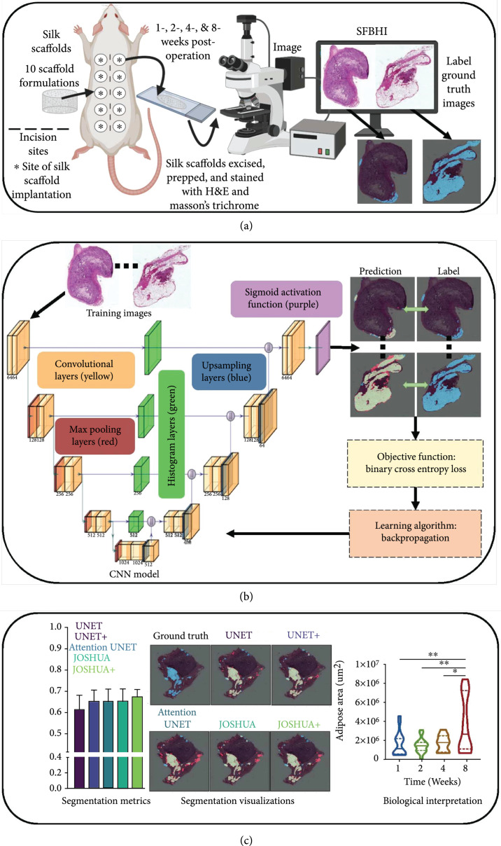
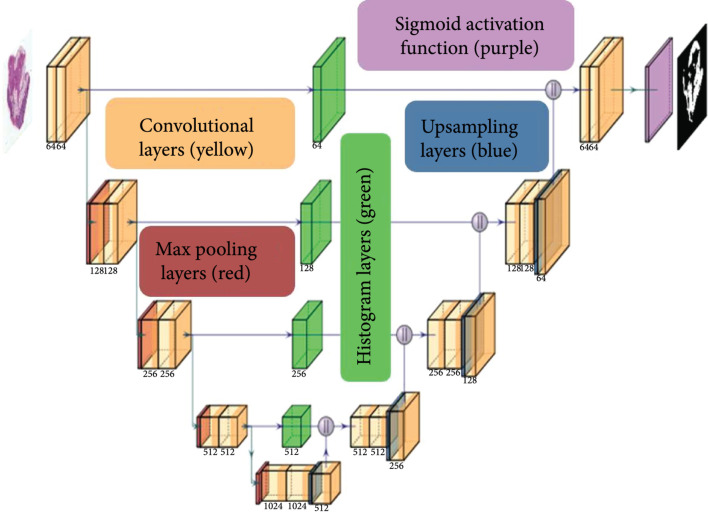
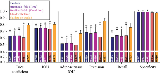
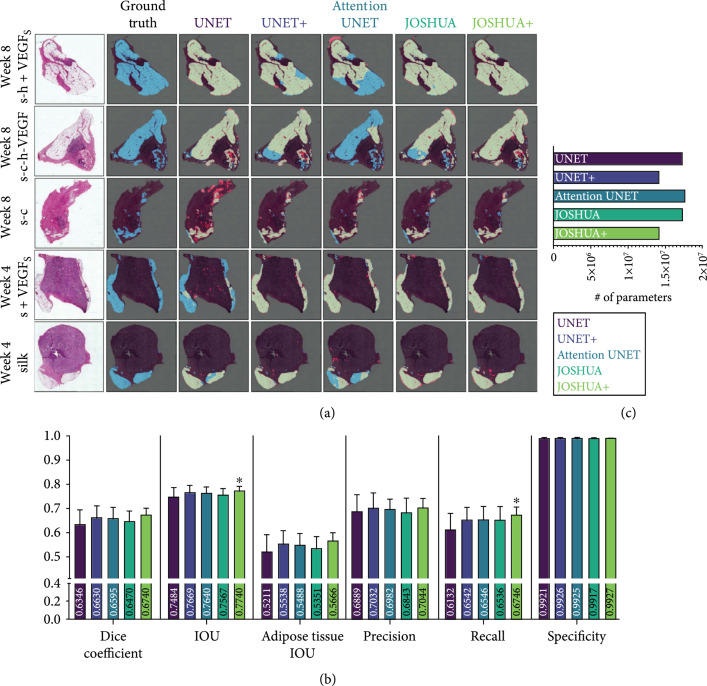
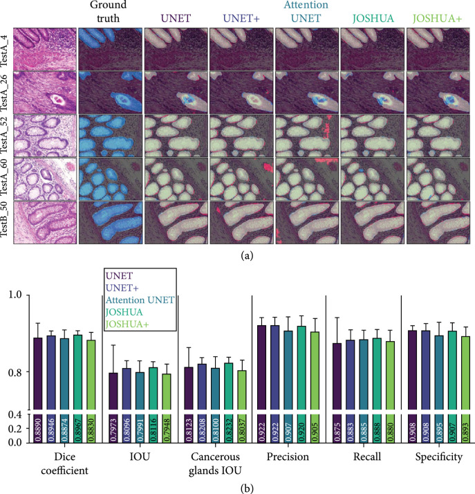
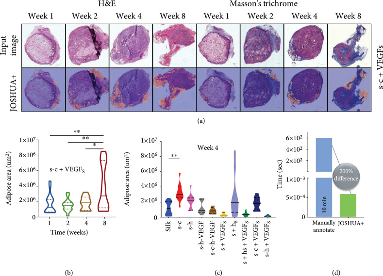
. We aim to develop a machine learning algorithm to quantify adipose tissue deposition at surgical sites as a function of biomaterial implantation. . To our knowledge, this study is the first investigation to apply convolutional neural network (CNN) models to identify and segment adipose tissue in histological images from silk fibroin biomaterial implants. . When designing biomaterials for the treatment of various soft tissue injuries and diseases, one must consider the extent of adipose tissue deposition. In this work, we analyzed adipose tissue accumulation in histological images of sectioned silk fibroin-based biomaterials excised from rodents following subcutaneous implantation for 1, 2, 4, or 8 weeks. Current strategies for quantifying adipose tissue after biomaterial implantation are often tedious and prone to human bias during analysis. . We used CNN models with novel spatial histogram layer(s) that can more accurately identify and segment regions of adipose tissue in hematoxylin and eosin (H&E) and Masson's trichrome stained images, allowing for determination of the optimal biomaterial formulation. We compared the method, Jointly Optimized Spatial Histogram UNET Architecture (JOSHUA), to the baseline UNET model and an extension of the baseline model, attention UNET, as well as to versions of the models with a supplemental attention-inspired mechanism (JOSHUA+ and UNET+). . The inclusion of histogram layer(s) in our models shows improved performance through qualitative and quantitative evaluation. . Our results demonstrate that the proposed methods, JOSHUA and JOSHUA+, are highly beneficial for adipose tissue identification and localization. The new histological dataset and code used in our experiments are publicly available.

摘要

我们旨在开发一种机器学习算法,以量化手术部位的脂肪组织沉积情况,作为生物材料植入的函数。据我们所知,本研究是首次将卷积神经网络(CNN)模型应用于识别和分割丝素蛋白生物材料植入物组织学图像中的脂肪组织。在设计用于治疗各种软组织损伤和疾病的生物材料时,必须考虑脂肪组织沉积的程度。在这项工作中,我们分析了从皮下植入1、2、4或8周后的啮齿动物身上切除的丝素蛋白基生物材料切片的组织学图像中的脂肪组织积累情况。目前生物材料植入后量化脂肪组织的策略通常很繁琐,且在分析过程中容易出现人为偏差。我们使用了带有新型空间直方图层的CNN模型,该模型能够更准确地识别和分割苏木精和伊红(H&E)以及Masson三色染色图像中的脂肪组织区域,从而确定最佳的生物材料配方。我们将联合优化空间直方图UNET架构(JOSHUA)方法与基线UNET模型、基线模型的扩展注意力UNET以及具有补充注意力启发机制的模型版本(JOSHUA+和UNET+)进行了比较。我们模型中包含直方图层通过定性和定量评估显示出了更好的性能。我们的结果表明,所提出的方法JOSHUA和JOSHUA+对脂肪组织的识别和定位非常有益。我们实验中使用的新组织学数据集和代码已公开可用。

https://cdn.ncbi.nlm.nih.gov/pmc/blobs/a52d/10521712/dbdbd8019949/9854084.fig.006.jpg
https://cdn.ncbi.nlm.nih.gov/pmc/blobs/a52d/10521712/2173b605bf35/9854084.fig.001.jpg
https://cdn.ncbi.nlm.nih.gov/pmc/blobs/a52d/10521712/d67750454460/9854084.fig.002.jpg
https://cdn.ncbi.nlm.nih.gov/pmc/blobs/a52d/10521712/c35696a2dde1/9854084.fig.003.jpg
https://cdn.ncbi.nlm.nih.gov/pmc/blobs/a52d/10521712/4bba24422637/9854084.fig.004.jpg
https://cdn.ncbi.nlm.nih.gov/pmc/blobs/a52d/10521712/d0ab051c6d84/9854084.fig.005.jpg
https://cdn.ncbi.nlm.nih.gov/pmc/blobs/a52d/10521712/dbdbd8019949/9854084.fig.006.jpg

相似文献

1
Jointly Optimized Spatial Histogram UNET Architecture (JOSHUA) for Adipose Tissue Segmentation.用于脂肪组织分割的联合优化空间直方图UNET架构(JOSHUA)
BME Front. 2022 Jun 3;2022:9854084. doi: 10.34133/2022/9854084. eCollection 2022.
2
Attention-VGG16-UNet: a novel deep learning approach for automatic segmentation of the median nerve in ultrasound images.注意力-VGG16-UNet:一种用于超声图像中正中神经自动分割的新型深度学习方法。
Quant Imaging Med Surg. 2022 Jun;12(6):3138-3150. doi: 10.21037/qims-21-1074.
3
Dense-UNet: a novel multiphoton cellular image segmentation model based on a convolutional neural network.密集型U-Net:一种基于卷积神经网络的新型多光子细胞图像分割模型。
Quant Imaging Med Surg. 2020 Jun;10(6):1275-1285. doi: 10.21037/qims-19-1090.
4
Deep learning-based recognition and segmentation of intracranial aneurysms under small sample size.小样本量下基于深度学习的颅内动脉瘤识别与分割
Front Physiol. 2022 Dec 19;13:1084202. doi: 10.3389/fphys.2022.1084202. eCollection 2022.
5
Efficient multiscale fully convolutional UNet model for segmentation of 3D lung nodule from CT image.用于从CT图像中分割3D肺结节的高效多尺度全卷积UNet模型。
J Med Imaging (Bellingham). 2022 Sep;9(5):052402. doi: 10.1117/1.JMI.9.5.052402. Epub 2022 May 11.
6
Automatic fetal biometry prediction using a novel deep convolutional network architecture.利用新型深度卷积网络架构进行自动胎儿生物测量预测。
Phys Med. 2021 Aug;88:127-137. doi: 10.1016/j.ejmp.2021.06.020. Epub 2021 Jul 6.
7
CTA-UNet: CNN-transformer architecture UNet for dental CBCT images segmentation.CTA-Unet:用于口腔 CBCT 图像分割的 CNN-Transformer 结构 U-Net。
Phys Med Biol. 2023 Aug 31;68(17). doi: 10.1088/1361-6560/acf026.
8
SW-UNet: a U-Net fusing sliding window transformer block with CNN for segmentation of lung nodules.SW-UNet:一种将滑动窗口变压器模块与卷积神经网络融合用于肺结节分割的U-Net。
Front Med (Lausanne). 2023 Sep 28;10:1273441. doi: 10.3389/fmed.2023.1273441. eCollection 2023.
9
A Medical Image Segmentation Method Based on Improved UNet 3+ Network.一种基于改进的UNet 3+网络的医学图像分割方法。
Diagnostics (Basel). 2023 Feb 3;13(3):576. doi: 10.3390/diagnostics13030576.
10
ConvWin-UNet: UNet-like hierarchical vision Transformer combined with convolution for medical image segmentation.卷积窗口U-Net:结合卷积的类U-Net层次视觉Transformer用于医学图像分割
Math Biosci Eng. 2023 Jan;20(1):128-144. doi: 10.3934/mbe.2023007. Epub 2022 Sep 29.

引用本文的文献

1
A Deep Learning-Based Automatic Segmentation and 3D Visualization Technique for Intracranial Hemorrhage Detection Using Computed Tomography Images.一种基于深度学习的自动分割与三维可视化技术,用于利用计算机断层扫描图像检测颅内出血。
Diagnostics (Basel). 2023 Jul 31;13(15):2537. doi: 10.3390/diagnostics13152537.
2
A strategy to quantify myofibroblast activation on a continuous spectrum.一种量化肌成纤维细胞激活的连续谱策略。
Sci Rep. 2022 Jul 18;12(1):12239. doi: 10.1038/s41598-022-16158-7.

本文引用的文献

1
Unified Focal loss: Generalising Dice and cross entropy-based losses to handle class imbalanced medical image segmentation.统一焦点损失:将基于 Dice 和交叉熵的损失函数推广到处理类不平衡的医学图像分割。
Comput Med Imaging Graph. 2022 Jan;95:102026. doi: 10.1016/j.compmedimag.2021.102026. Epub 2021 Dec 13.
2
Deep Interpretable Classification and Weakly-Supervised Segmentation of Histology Images via Max-Min Uncertainty.基于极大极小不确定性的组织学图像深度可解释分类与弱监督分割。
IEEE Trans Med Imaging. 2022 Mar;41(3):702-714. doi: 10.1109/TMI.2021.3123461. Epub 2022 Mar 2.
3
Texture Analysis and Its Applications in Biomedical Imaging: A Survey.
纹理分析及其在生物医学成像中的应用:综述。
IEEE Rev Biomed Eng. 2022;15:222-246. doi: 10.1109/RBME.2021.3115703. Epub 2022 Jan 20.
4
Image Segmentation Using Deep Learning: A Survey.基于深度学习的图像分割技术综述。
IEEE Trans Pattern Anal Mach Intell. 2022 Jul;44(7):3523-3542. doi: 10.1109/TPAMI.2021.3059968. Epub 2022 Jun 3.
5
Analyzing Overfitting Under Class Imbalance in Neural Networks for Image Segmentation.分析图像分割中神经网络的类别不平衡过拟合问题。
IEEE Trans Med Imaging. 2021 Mar;40(3):1065-1077. doi: 10.1109/TMI.2020.3046692. Epub 2021 Mar 2.
6
Deep neural network models for computational histopathology: A survey.用于计算组织病理学的深度神经网络模型:一项综述。
Med Image Anal. 2021 Jan;67:101813. doi: 10.1016/j.media.2020.101813. Epub 2020 Sep 25.
7
UNet++: A Nested U-Net Architecture for Medical Image Segmentation.U-Net++:一种用于医学图像分割的嵌套U-Net架构。
Deep Learn Med Image Anal Multimodal Learn Clin Decis Support (2018). 2018 Sep;11045:3-11. doi: 10.1007/978-3-030-00889-5_1. Epub 2018 Sep 20.
8
Automated segmentation of abdominal organs from contrast-enhanced computed tomography using analysis of texture features.基于纹理特征分析的增强 CT 腹部器官自动分割。
Int J Numer Method Biomed Eng. 2020 Apr;36(4):e3309. doi: 10.1002/cnm.3309. Epub 2020 Mar 3.
9
Silk fibroin for skin injury repair: Where do things stand?丝素蛋白在皮肤损伤修复中的应用:现状如何?
Adv Drug Deliv Rev. 2020 Jan 1;153:28-53. doi: 10.1016/j.addr.2019.09.003. Epub 2019 Oct 31.
10
Asymmetric Loss Functions and Deep Densely Connected Networks for Highly Imbalanced Medical Image Segmentation: Application to Multiple Sclerosis Lesion Detection.用于高度不平衡医学图像分割的非对称损失函数和深度密集连接网络:在多发性硬化症病变检测中的应用
IEEE Access. 2019;7:721-1735. doi: 10.1109/ACCESS.2018.2886371. Epub 2018 Dec 12.