• 文献检索
  • 文档翻译
  • 深度研究
  • 学术资讯
  • Suppr Zotero 插件Zotero 插件
  • 邀请有礼
  • 套餐&价格
  • 历史记录
应用&插件
Suppr Zotero 插件Zotero 插件浏览器插件Mac 客户端Windows 客户端微信小程序
定价
高级版会员购买积分包购买API积分包
服务
文献检索文档翻译深度研究API 文档MCP 服务
关于我们
关于 Suppr公司介绍联系我们用户协议隐私条款
关注我们

Suppr 超能文献

核心技术专利:CN118964589B侵权必究
粤ICP备2023148730 号-1Suppr @ 2026

文献检索

告别复杂PubMed语法,用中文像聊天一样搜索,搜遍4000万医学文献。AI智能推荐,让科研检索更轻松。

立即免费搜索

文件翻译

保留排版,准确专业,支持PDF/Word/PPT等文件格式,支持 12+语言互译。

免费翻译文档

深度研究

AI帮你快速写综述,25分钟生成高质量综述,智能提取关键信息,辅助科研写作。

立即免费体验

通过跨模态预训练实现一次性视网膜动静脉分割

One-shot Retinal Artery and Vein Segmentation via Cross-modality Pretraining.

作者信息

Shi Danli, He Shuang, Yang Jiancheng, Zheng Yingfeng, He Mingguang

机构信息

Centre for Eye and Vision Research (CEVR), Hong Kong SAR, China.

The Hong Kong Polytechnic University, Kowloon, Hong Kong SAR, China.

出版信息

Ophthalmol Sci. 2023 Jul 6;4(2):100363. doi: 10.1016/j.xops.2023.100363. eCollection 2024 Mar-Apr.

DOI:10.1016/j.xops.2023.100363
PMID:37868792
原文链接:https://pmc.ncbi.nlm.nih.gov/articles/PMC10585631/
Abstract

PURPOSE

To perform one-shot retinal artery and vein segmentation with cross-modality artery-vein (AV) soft-label pretraining.

DESIGN

Cross-sectional study.

SUBJECTS

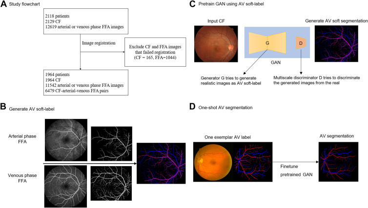
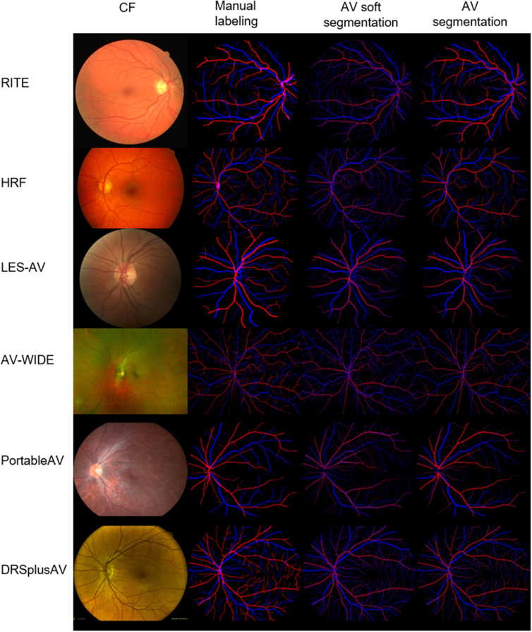
The study included 6479 color fundus photography (CFP) and arterial-venous fundus fluorescein angiography (FFA) pairs from 1964 participants for pretraining and 6 AV segmentation data sets with various image sources, including RITE, HRF, LES-AV, AV-WIDE, PortableAV, and DRSplusAV for one-shot finetuning and testing.

METHODS

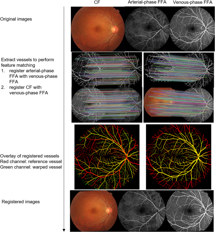
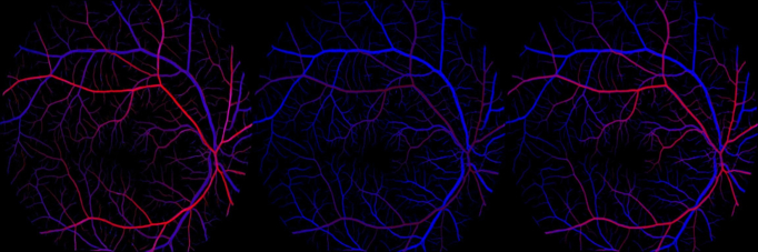
We structurally matched the arterial and venous phase of FFA with CFP, the AV soft labels were automatically generated by utilizing the fluorescein intensity difference of the arterial and venous-phase FFA images, and the soft labels were then used to train a generative adversarial network to learn to generate AV soft segmentations using CFP images as input. We then finetuned the pretrained model to perform AV segmentation using only one image from each of the AV segmentation data sets and test on the remainder. To investigate the effect and reliability of one-shot finetuning, we conducted experiments without finetuning and by finetuning the pretrained model on an iteratively different single image for each data set under the same experimental setting and tested the models on the remaining images.

MAIN OUTCOME MEASURES

The AV segmentation was assessed by area under the receiver operating characteristic curve (AUC), accuracy, Dice score, sensitivity, and specificity.

RESULTS

After the FFA-AV soft label pretraining, our method required only one exemplar image from each camera or modality and achieved similar performance with full-data training, with AUC ranging from 0.901 to 0.971, accuracy from 0.959 to 0.980, Dice score from 0.585 to 0.773, sensitivity from 0.574 to 0.763, and specificity from 0.981 to 0.991. Compared with no finetuning, the segmentation performance improved after one-shot finetuning. When finetuned on different images in each data set, the standard deviation of the segmentation results across models ranged from 0.001 to 0.10.

CONCLUSIONS

This study presents the first one-shot approach to retinal artery and vein segmentation. The proposed labeling method is time-saving and efficient, demonstrating a promising direction for retinal-vessel segmentation and enabling the potential for widespread application.

FINANCIAL DISCLOSURES

Proprietary or commercial disclosure may be found in the Footnotes and Disclosures at the end of this article.

摘要

目的

通过跨模态动静脉(AV)软标签预训练进行一次性视网膜动静脉分割。

设计

横断面研究。

研究对象

该研究纳入了来自1964名参与者的6479对彩色眼底照片(CFP)和动静脉眼底荧光血管造影(FFA)用于预训练,以及6个具有不同图像来源的AV分割数据集,包括RITE、HRF、LES-AV、AV-WIDE、PortableAV和DRSplusAV用于一次性微调及测试。

方法

我们在结构上使FFA的动静脉期与CFP匹配,利用动静脉期FFA图像的荧光素强度差异自动生成AV软标签,然后使用这些软标签训练一个生成对抗网络,以学习使用CFP图像作为输入来生成AV软分割。然后,我们对预训练模型进行微调,以便仅使用每个AV分割数据集中的一张图像来执行AV分割,并在其余图像上进行测试。为了研究一次性微调的效果和可靠性,我们在相同实验设置下,进行了不微调以及在每个数据集的迭代不同单张图像上对预训练模型进行微调的实验,并在其余图像上测试模型。

主要观察指标

通过受试者操作特征曲线下面积(AUC)、准确率、Dice系数、灵敏度和特异性来评估AV分割。

结果

经过FFA-AV软标签预训练后,我们的方法每个相机或模态仅需一张示例图像,且在全数据训练下取得了相似的性能,AUC范围为0.901至0.971,准确率为0.959至0.980,Dice系数为0.585至0.773,灵敏度为0.574至0.763,特异性为0.981至0.991。与不微调相比,一次性微调后分割性能有所提高。当在每个数据集中的不同图像上进行微调时,各模型分割结果的标准差范围为0.001至0.10。

结论

本研究提出了首个用于视网膜动静脉分割的一次性方法。所提出的标记方法省时高效为视网膜血管分割展示了一个有前景的方向,并具有广泛应用的潜力。

财务披露

在本文末尾的脚注和披露中可能会找到专有或商业披露信息。

https://cdn.ncbi.nlm.nih.gov/pmc/blobs/a26f/10585631/f53813ce6ea2/gr4.jpg
https://cdn.ncbi.nlm.nih.gov/pmc/blobs/a26f/10585631/ca286e09a58b/gr1.jpg
https://cdn.ncbi.nlm.nih.gov/pmc/blobs/a26f/10585631/9d7e1624fb2a/gr2.jpg
https://cdn.ncbi.nlm.nih.gov/pmc/blobs/a26f/10585631/09cc478fa486/gr3.jpg
https://cdn.ncbi.nlm.nih.gov/pmc/blobs/a26f/10585631/f53813ce6ea2/gr4.jpg
https://cdn.ncbi.nlm.nih.gov/pmc/blobs/a26f/10585631/ca286e09a58b/gr1.jpg
https://cdn.ncbi.nlm.nih.gov/pmc/blobs/a26f/10585631/9d7e1624fb2a/gr2.jpg
https://cdn.ncbi.nlm.nih.gov/pmc/blobs/a26f/10585631/09cc478fa486/gr3.jpg
https://cdn.ncbi.nlm.nih.gov/pmc/blobs/a26f/10585631/f53813ce6ea2/gr4.jpg

相似文献

1
One-shot Retinal Artery and Vein Segmentation via Cross-modality Pretraining.通过跨模态预训练实现一次性视网膜动静脉分割
Ophthalmol Sci. 2023 Jul 6;4(2):100363. doi: 10.1016/j.xops.2023.100363. eCollection 2024 Mar-Apr.
2
Cross-modality Labeling Enables Noninvasive Capillary Quantification as a Sensitive Biomarker for Assessing Cardiovascular Risk.跨模态标记实现无创性毛细血管定量分析,作为评估心血管风险的敏感生物标志物。
Ophthalmol Sci. 2023 Dec 5;4(3):100441. doi: 10.1016/j.xops.2023.100441. eCollection 2024 May-Jun.
3
Translation of Color Fundus Photography into Fluorescein Angiography Using Deep Learning for Enhanced Diabetic Retinopathy Screening.利用深度学习将彩色眼底照片转换为荧光血管造影以增强糖尿病视网膜病变筛查
Ophthalmol Sci. 2023 Sep 15;3(4):100401. doi: 10.1016/j.xops.2023.100401. eCollection 2023 Dec.
4
Lesion-aware generative adversarial networks for color fundus image to fundus fluorescein angiography translation.用于彩色眼底图像到眼底荧光血管造影转换的病变感知生成对抗网络。
Comput Methods Programs Biomed. 2023 Feb;229:107306. doi: 10.1016/j.cmpb.2022.107306. Epub 2022 Dec 14.
5
Multi-path cascaded U-net for vessel segmentation from fundus fluorescein angiography sequential images.多路径级联 U-net 用于从眼底荧光素血管造影序列图像中进行血管分割。
Comput Methods Programs Biomed. 2021 Nov;211:106422. doi: 10.1016/j.cmpb.2021.106422. Epub 2021 Sep 20.
6
Artery-vein segmentation in fundus images using a fully convolutional network.基于全卷积网络的眼底图像动静脉分割。
Comput Med Imaging Graph. 2019 Sep;76:101636. doi: 10.1016/j.compmedimag.2019.05.004. Epub 2019 Jun 15.
7
Low-Shot Deep Learning of Diabetic Retinopathy With Potential Applications to Address Artificial Intelligence Bias in Retinal Diagnostics and Rare Ophthalmic Diseases.基于少量样本的深度学习在糖尿病视网膜病变中的应用及其对解决视网膜诊断中人工智能偏倚和罕见眼病问题的潜力。
JAMA Ophthalmol. 2020 Oct 1;138(10):1070-1077. doi: 10.1001/jamaophthalmol.2020.3269.
8
[A new approach for studying the retinal and choroidal circulation].[一种研究视网膜和脉络膜循环的新方法]
Nippon Ganka Gakkai Zasshi. 2004 Dec;108(12):836-61; discussion 862.
9
Artery vein classification in fundus images using serially connected U-Nets.基于连续 U-Net 的眼底图像动静脉分类。
Comput Methods Programs Biomed. 2022 Apr;216:106650. doi: 10.1016/j.cmpb.2022.106650. Epub 2022 Jan 23.
10
NCCT-CECT image synthesizers and their application to pulmonary vessel segmentation.非增强计算机断层扫描-增强计算机断层扫描图像合成器及其在肺血管分割中的应用
Comput Methods Programs Biomed. 2023 Apr;231:107389. doi: 10.1016/j.cmpb.2023.107389. Epub 2023 Feb 2.

引用本文的文献

1
Generation of Fundus Fluorescein Angiography Videos for Health Care Data Sharing.用于医疗保健数据共享的眼底荧光血管造影视频生成
JAMA Ophthalmol. 2025 Jun 26. doi: 10.1001/jamaophthalmol.2025.1419.
2
A multimodal visual-language foundation model for computational ophthalmology.一种用于计算机眼科的多模态视觉语言基础模型。
NPJ Digit Med. 2025 Jun 21;8(1):381. doi: 10.1038/s41746-025-01772-2.
3
A Future Picture: A Review of Current Generative Adversarial Neural Networks in Vitreoretinal Pathologies and Their Future Potentials.

本文引用的文献

1
A Deep Learning System for Fully Automated Retinal Vessel Measurement in High Throughput Image Analysis.一种用于高通量图像分析中全自动视网膜血管测量的深度学习系统。
Front Cardiovasc Med. 2022 Mar 22;9:823436. doi: 10.3389/fcvm.2022.823436. eCollection 2022.
2
Artery vein classification in fundus images using serially connected U-Nets.基于连续 U-Net 的眼底图像动静脉分类。
Comput Methods Programs Biomed. 2022 Apr;216:106650. doi: 10.1016/j.cmpb.2022.106650. Epub 2022 Jan 23.
3
TW-GAN: Topology and width aware GAN for retinal artery/vein classification.
未来图景:当前生成对抗神经网络在玻璃体视网膜病变中的应用及其未来潜力综述
Biomedicines. 2025 Jan 24;13(2):284. doi: 10.3390/biomedicines13020284.
4
ChatFFA: An ophthalmic chat system for unified vision-language understanding and question answering for fundus fluorescein angiography.ChatFFA:一种用于眼底荧光血管造影的统一视觉语言理解和问答的眼科聊天系统。
iScience. 2024 May 17;27(7):110021. doi: 10.1016/j.isci.2024.110021. eCollection 2024 Jul 19.
5
LiViT-Net: A U-Net-like, lightweight Transformer network for retinal vessel segmentation.LiViT-Net:一种用于视网膜血管分割的类似U-Net的轻量级Transformer网络。
Comput Struct Biotechnol J. 2024 Mar 19;24:213-224. doi: 10.1016/j.csbj.2024.03.003. eCollection 2024 Dec.
6
Cross-modality Labeling Enables Noninvasive Capillary Quantification as a Sensitive Biomarker for Assessing Cardiovascular Risk.跨模态标记实现无创性毛细血管定量分析,作为评估心血管风险的敏感生物标志物。
Ophthalmol Sci. 2023 Dec 5;4(3):100441. doi: 10.1016/j.xops.2023.100441. eCollection 2024 May-Jun.
7
Translating color fundus photography to indocyanine green angiography using deep-learning for age-related macular degeneration screening.利用深度学习将彩色眼底摄影转换为吲哚菁绿血管造影以进行年龄相关性黄斑变性筛查。
NPJ Digit Med. 2024 Feb 12;7(1):34. doi: 10.1038/s41746-024-01018-7.
8
Bridging the Camera Domain Gap With Image-to-Image Translation Improves Glaucoma Diagnosis.通过图像到图像的翻译弥合相机域差距可提高青光眼诊断。
Transl Vis Sci Technol. 2023 Dec 1;12(12):20. doi: 10.1167/tvst.12.12.20.
9
A deep learning model for generating fundus autofluorescence images from color fundus photography.一种用于从彩色眼底照片生成眼底自发荧光图像的深度学习模型。
Adv Ophthalmol Pract Res. 2023 Nov 10;3(4):192-198. doi: 10.1016/j.aopr.2023.11.001. eCollection 2023 Nov-Dec.
10
Translation of Color Fundus Photography into Fluorescein Angiography Using Deep Learning for Enhanced Diabetic Retinopathy Screening.利用深度学习将彩色眼底照片转换为荧光血管造影以增强糖尿病视网膜病变筛查
Ophthalmol Sci. 2023 Sep 15;3(4):100401. doi: 10.1016/j.xops.2023.100401. eCollection 2023 Dec.
TW-GAN:用于视网膜动脉/静脉分类的拓扑和宽度感知 GAN。
Med Image Anal. 2022 Apr;77:102340. doi: 10.1016/j.media.2021.102340. Epub 2021 Dec 23.
4
Simultaneous segmentation and classification of the retinal arteries and veins from color fundus images.彩色眼底图像中视网膜动脉和静脉的同时分割和分类。
Artif Intell Med. 2021 Aug;118:102116. doi: 10.1016/j.artmed.2021.102116. Epub 2021 May 29.
5
Imaging the Retinal Vasculature.视网膜血管成像。
Annu Rev Vis Sci. 2021 Sep 15;7:129-153. doi: 10.1146/annurev-vision-093019-113719. Epub 2021 Jun 25.
6
A global review of publicly available datasets for ophthalmological imaging: barriers to access, usability, and generalisability.眼科成像公共可用数据集的全球回顾:获取、可用性和可推广性的障碍。
Lancet Digit Health. 2021 Jan;3(1):e51-e66. doi: 10.1016/S2589-7500(20)30240-5. Epub 2020 Oct 1.
7
A review of machine learning methods for retinal blood vessel segmentation and artery/vein classification.机器学习方法在视网膜血管分割和动静脉分类中的研究进展综述。
Med Image Anal. 2021 Feb;68:101905. doi: 10.1016/j.media.2020.101905. Epub 2020 Nov 17.
8
A deep-learning system for the assessment of cardiovascular disease risk via the measurement of retinal-vessel calibre.一种通过测量视网膜血管口径评估心血管疾病风险的深度学习系统。
Nat Biomed Eng. 2021 Jun;5(6):498-508. doi: 10.1038/s41551-020-00626-4. Epub 2020 Oct 12.
9
Retinal biomarkers for Alzheimer's disease and vascular cognitive impairment and dementia (VCID): implication for early diagnosis and prognosis.阿尔茨海默病和血管性认知障碍及痴呆(VCID)的视网膜生物标志物:对早期诊断和预后的影响。
Geroscience. 2020 Dec;42(6):1499-1525. doi: 10.1007/s11357-020-00252-7. Epub 2020 Oct 4.
10
AVNet: A retinal artery/vein classification network with category-attention weighted fusion.AVNet:一种具有类别注意力加权融合的视网膜动脉/静脉分类网络。
Comput Methods Programs Biomed. 2020 Oct;195:105629. doi: 10.1016/j.cmpb.2020.105629. Epub 2020 Jun 25.