Department of Nutritional Sciences, College of Agricultural & Life Sciences, University of Wisconsin, Madison, Wisconsin, USA.
Department of Ophthalmology and Visual Sciences, University of Wisconsin, Madison, Wisconsin, USA.
BMJ Open Ophthalmol. 2023 Oct;8(1). doi: 10.1136/bmjophth-2023-001331.
To investigate associations between baseline macular pigment optical density (MPOD) and retinal layer thicknesses in eyes with and without manifest primary open-angle glaucoma (POAG) in the Carotenoids in Age-Related Eye Disease Study 2 (CAREDS2).
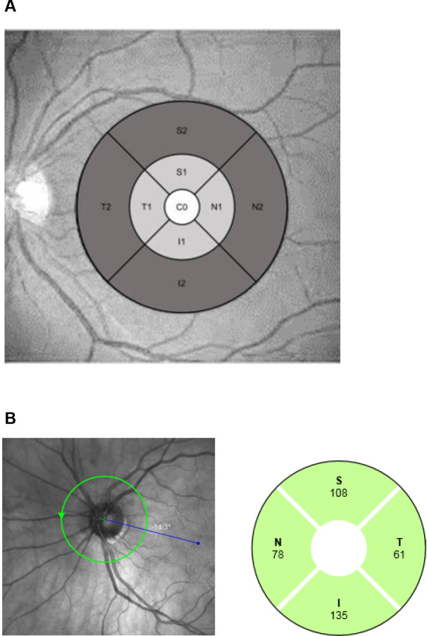
MPOD was measured at CAREDS baseline (2001-2004) via heterochromatic flicker photometry (0.5° from foveal centre). Peripapillary retinal nerve fibre layer (RNFL), macular ganglion cell complex (GCC), ganglion cell layer (GCL), inner plexiform layer (IPL), and RNFL thicknesses were measured at CAREDS2 (2016-2019) via spectral-domain optical coherence tomography. Associations between MPOD and retinal thickness were assessed using multivariable linear regression.
Among 742 eyes (379 participants), manifest POAG was identified in 50 eyes (32 participants). In eyes without manifest POAG, MPOD was positively associated with macular GCC, GCL and IPL thicknesses in the central subfield (P-trend ≤0.01), but not the inner or outer subfields. Among eyes with manifest POAG, MPOD was positively associated with macular GCC, GCL, IPL and RNFL in the central subfield (P-trend ≤0.03), but not the inner or outer subfields, and was positively associated with peripapillary RNFL thickness in the superior and temporal quadrants (P-trend≤0.006).
We observed a positive association between MPOD and central subfield GCC thickness 15 years later. MPOD was positively associated with peripapillary RNFL superior and temporal quadrant thicknesses among eyes with manifest POAG. Our results linking low MPOD to retinal layers that are structural indicators of early glaucoma provide further evidence that carotenoids may be protective against manifest POAG.
在 Carotenoids in Age-Related Eye Disease Study 2(CAREDS2)中,调查具有和不具有明显原发性开角型青光眼(POAG)的眼的基线黄斑色素光学密度(MPOD)与视网膜层厚度之间的关联。
通过异色调闪烁光度计(距中心凹 0.5°)在 CAREDS 基线(2001-2004 年)测量 MPOD。通过光谱域光学相干断层扫描在 CAREDS2(2016-2019 年)测量周边视网膜神经纤维层(RNFL)、黄斑神经节细胞复合体(GCC)、神经节细胞层(GCL)、内丛状层(IPL)和 RNFL 厚度。使用多变量线性回归评估 MPOD 与视网膜厚度之间的关联。
在 742 只眼(379 名参与者)中,确定了 50 只眼(32 名参与者)患有明显 POAG。在没有明显 POAG 的眼中,MPOD 与中央凹的黄斑 GCC、GCL 和 IPL 厚度呈正相关(P 趋势≤0.01),但与内或外凹无相关性。在具有明显 POAG 的眼中,MPOD 与中央凹的黄斑 GCC、GCL、IPL 和 RNFL 呈正相关(P 趋势≤0.03),但与内或外凹无相关性,并且与上方和颞象限的周边 RNFL 厚度呈正相关(P 趋势≤0.006)。
我们观察到 15 年后 MPOD 与中央凹 GCC 厚度之间存在正相关。在具有明显 POAG 的眼中,MPOD 与上方和颞象限的周边 RNFL 厚度呈正相关。我们将低 MPOD 与作为早期青光眼结构指标的视网膜层联系起来的结果提供了进一步的证据,表明类胡萝卜素可能对明显 POAG 具有保护作用。