文献检索文档翻译深度研究
Suppr Zotero 插件Zotero 插件
邀请有礼套餐&价格历史记录

新学期,新优惠

限时优惠:9月1日-9月22日

30天高级会员仅需29元

1天体验卡首发特惠仅需5.99元

了解详情
不再提醒
插件&应用
Suppr Zotero 插件Zotero 插件浏览器插件Mac 客户端Windows 客户端微信小程序
高级版
套餐订阅购买积分包
AI 工具
文献检索文档翻译深度研究
关于我们
关于 Suppr公司介绍联系我们用户协议隐私条款
关注我们

Suppr 超能文献

核心技术专利:CN118964589B侵权必究
粤ICP备2023148730 号-1Suppr @ 2025

Longitudinal Three-Dimensional Stereophotogrammetric Growth Analysis in Infants with Unilateral Cleft Lip and Palate from 3 to 12 Months of Age.

作者信息

Kluge Jennifer, Bruggink Robin, Pandis Nikolaos, Unkovskiy Alexey, Jost-Brinkmann Paul-Georg, Kuijpers-Jagtman Anne Marie, Bartzela Theodosia

机构信息

Department of Orthodontics and Dentofacial Orthopedics, Center for Oral Health Sciences CC3, Charité-Universitätsmedizin Berlin, Corporate Member of Freie Universität Berlin and Humboldt-Universität zu Berlin, Aßmannshauser Straße 4-6, 14197 Berlin, Germany.

Radboudumc 3D Lab, Radboud Institute for Health Sciences, Radboud University Medical Center, 6500 HB Nijmegen, The Netherlands.

出版信息

J Clin Med. 2023 Oct 10;12(20):6432. doi: 10.3390/jcm12206432.


DOI:10.3390/jcm12206432
PMID:37892569
原文链接:https://pmc.ncbi.nlm.nih.gov/articles/PMC10607132/
Abstract

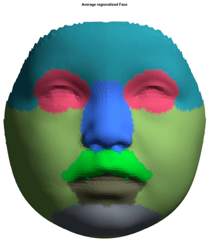
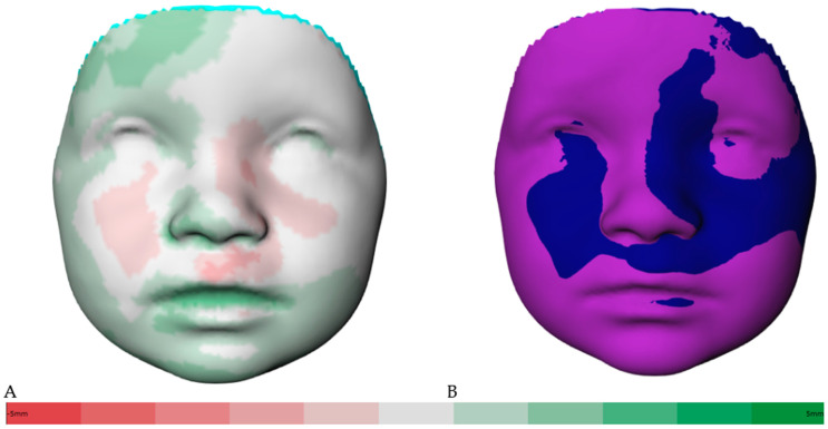
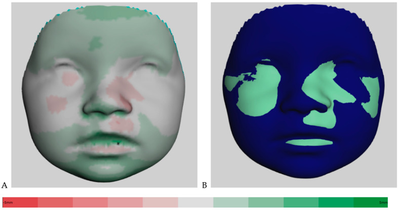
This longitudinal study aimed to evaluate facial growth and soft tissue changes in infants with complete unilateral cleft lip, alveolus, and palate (CUCLAP) at ages 3, 9, and 12 months. Using 3D images of 22 CUCLAP infants, average faces and distance maps for the entire face and specific regions were created. Color-coded maps highlighted more significant soft tissue changes from 3 to 9 months than from 9 to 12 months. The first interval showed substantial growth in the entire face, particularly in the forehead, eyes, lower lip, chin, and cheeks ( < 0.001), while the second interval exhibited no significant growth. This study provides insights into facial soft tissue growth in CUCLAP infants during critical developmental stages, emphasizing substantial improvements between 3 and 9 months, mainly in the chin, lower lip, and forehead. However, uneven growth occurred in the upper lip, philtrum, and nostrils throughout both intervals, with an overall decline in growth from 9 to 12 months. These findings underscore the dynamic nature of soft tissue growth in CUCLAP patients, highlighting the need to consider these patterns in treatment planning. Future research should explore the underlying factors and develop customized treatment interventions for enhanced facial aesthetics and function in this population.

摘要
https://cdn.ncbi.nlm.nih.gov/pmc/blobs/ff24/10607132/062ca9e78605/jcm-12-06432-g003.jpg
https://cdn.ncbi.nlm.nih.gov/pmc/blobs/ff24/10607132/ac8b9bb76755/jcm-12-06432-g001.jpg
https://cdn.ncbi.nlm.nih.gov/pmc/blobs/ff24/10607132/26e16ffa60a8/jcm-12-06432-g002.jpg
https://cdn.ncbi.nlm.nih.gov/pmc/blobs/ff24/10607132/062ca9e78605/jcm-12-06432-g003.jpg
https://cdn.ncbi.nlm.nih.gov/pmc/blobs/ff24/10607132/ac8b9bb76755/jcm-12-06432-g001.jpg
https://cdn.ncbi.nlm.nih.gov/pmc/blobs/ff24/10607132/26e16ffa60a8/jcm-12-06432-g002.jpg
https://cdn.ncbi.nlm.nih.gov/pmc/blobs/ff24/10607132/062ca9e78605/jcm-12-06432-g003.jpg

相似文献

[1]
Longitudinal Three-Dimensional Stereophotogrammetric Growth Analysis in Infants with Unilateral Cleft Lip and Palate from 3 to 12 Months of Age.

J Clin Med. 2023-10-10

[2]
Three-dimensional facial development of children with unilateral cleft lip and palate during the first year of life in comparison with normative average faces.

PeerJ. 2019-7-30

[3]
Assessment of regional asymmetry of the face before and after surgical correction of unilateral cleft lip.

J Craniomaxillofac Surg. 2018-4-6

[4]
Longitudinal 3D Assessment of Facial Asymmetry in Unilateral Cleft Lip and Palate.

Cleft Palate Craniofac J. 2019-4

[5]
A Pilot Study on the Influence of Facial Expression on Measurements in Three-Dimensional Digital Surfaces of the Face in Infants With Cleft Lip and Palate.

Cleft Palate Craniofac J. 2016-1

[6]
Three-Dimensional Cone Beam Computed Tomography (CBCT)-Derived Soft Tissue Changes in Patients with Unilateral Cleft Lip, Alveolus, and Palate with Midfacial Deficiency after 1.5 Years of Bone-Anchored Maxillary Protraction.

J Clin Med. 2024-5-14

[7]
3D soft tissue changes in facial morphology in patients with cleft lip and palate and class III mal occlusion under therapy with rapid maxillary expansion and delaire facemask.

J Orofac Orthop. 2010-3

[8]
Three-Dimensional Soft-Tissue Evaluation in Patients with Cleft Lip and Palate.

Med Sci Monit. 2018-11-28

[9]
Regional facial asymmetries in unilateral orofacial clefts.

Eur J Orthod. 2015-12

[10]
Uniform 3D meshes to establish normative facial averages of healthy infants during the first year of life.

PLoS One. 2019-5-20

引用本文的文献

[1]
Three-dimensional analysis of facial morphology in nine-year-old children with different unilateral orofacial clefts compared to normative data.

PeerJ. 2025-1-21

本文引用的文献

[1]
Facial scanning technologies in the era of digital workflow: A systematic review and network meta-analysis.

J Prosthodont Res. 2023-7-31

[2]
A semi-automatic three-dimensional technique using a regionalized facial template enables facial growth assessment in healthy children from 1.5 to 5.0 years of age.

PeerJ. 2022

[3]
Effects of Artificial Extraoral Markers on Accuracy of Three-Dimensional Dentofacial Image Integration: Smartphone Face Scan versus Stereophotogrammetry.

J Pers Med. 2022-3-18

[4]
Large-scale open-source three-dimensional growth curves for clinical facial assessment and objective description of facial dysmorphism.

Sci Rep. 2021-6-9

[5]
Clinical characterization of 266 patients and family members with cleft lip and/or palate with associated malformations and syndromes.

Clin Oral Investig. 2021-9

[6]
Three-Dimensional Analysis of Craniofacial Structures of Individuals With Nonsyndromic Unilateral Complete Cleft Lip and Palate.

J Craniofac Surg.

[7]
Nasolabial shape and aesthetics in unilateral cleft lip and palate: an analysis of nasolabial shape using a mean 3D facial template.

Int J Oral Maxillofac Surg. 2021-2

[8]
Symmetry of palatal shape during the first year of life in healthy infants.

Clin Oral Investig. 2021-3

[9]
An Innovative Assessment of the Dynamics of Facial Movements in Surgically Managed Unilateral Cleft Lip and Palate Using 4D Imaging.

Cleft Palate Craniofac J. 2020-9

[10]
Three-dimensional facial development of children with unilateral cleft lip and palate during the first year of life in comparison with normative average faces.

PeerJ. 2019-7-30

文献AI研究员

20分钟写一篇综述,助力文献阅读效率提升50倍

立即体验

用中文搜PubMed

大模型驱动的PubMed中文搜索引擎

马上搜索

推荐工具

医学文档翻译智能文献检索