Department of Microbiology and Immunology, Faculty of Pharmacy, Helwan University, Ain Helwan, Cairo, Egypt.
Department of Microbiology and Immunology, Faculty of Pharmacy, Modern University for Technology and Information (MTI), Cairo, Egypt.
Braz J Microbiol. 2024 Mar;55(1):297-308. doi: 10.1007/s42770-023-01177-x. Epub 2023 Nov 18.
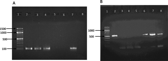
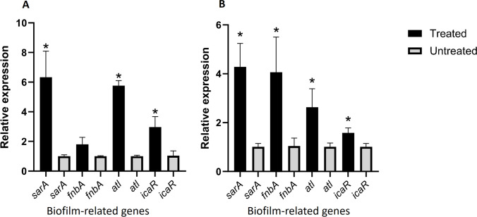
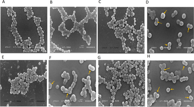
The exposure of bacteria to sub-inhibitory concentrations of antibiotics is of biological significance since it can occur in vivo under many circumstances, including low-dose treatment, poor adherence to a regimen, poor drug penetration, drug-drug interactions, and antibiotic resistance of the pathogen. In this study, we investigated the effects of subinhibitory concentrations of four antibiotics: ampicillin, ceftriaxone, gentamicin, and norfloxacin, which are commonly used in clinical settings and on cell morphology and biofilm formation in Staphylococcus aureus as one of the leading causes of nosocomial and biofilm-associated infections. Nine clinical S. aureus biofilm-producing isolates and two known biofilm-producing reference strains, S. aureus ATCC 29213 and S. aureus ATCC 6538, were used in this study. Sub-MICs of beta-lactam antibiotics (ampicillin and ceftriaxone) significantly induced biofilm formation in S. aureus ATCC 29213 and S. aureus ATCC 6538 and in six clinical isolates out of the nine selected isolates when compared with the antibiotic-free control group (P < 0.05), with an approximately 2- to 2.5-fold increase. Gentamicin and norfloxacin induced biofilms in S. aureus ATCC 29213 and S. aureus ATCC 6538, while gentamicin and norfloxacin induced biofilms only in three and two of the nine tested isolates, respectively (P < 0.05). The chemical nature of the biofilm matrix produced by half the MIC of ceftriaxone in the six isolates that showed increased biofilm was all non-polysaccharide in composition (PIA-independent). Gene expression of biofilm-encoding genes atl and sarA in biofilms of the two tested strains (S. aureus ATCC 6538) and clinical strain (S. aureus 16) showed a significant upregulation after exposure to half MIC of ceftriaxone. Additionally, the bacterial cell morphological changes in planktonic cells caused by half MIC of ceftriaxone were evaluated by scanning electron microscopy, which demonstrated a significant cell enlargement when compared with the antibiotic-free control (P < 0.05), and some deformed cells were also noticed. In S. aureus clinical isolates, sub-MICs of ampicillin, ceftriaxone, gentamicin, and norfloxacin may stimulate substantial production of biofilm, which could have important clinical significance and make infection treatment challenges. Further, in vivo research is needed to fully comprehend how sub-MIC of antibiotics can affect biofilm formation in clinical settings. Additionally, more research is required to reveal the clinical implications of the morphological alterations in S. aureus brought on by exposure to ceftriaxone at concentrations below its MIC.
细菌暴露于亚抑菌浓度的抗生素具有生物学意义,因为在许多情况下,包括低剂量治疗、治疗方案依从性差、药物渗透不良、药物相互作用和病原体的抗生素耐药性,这种情况可能在体内发生。在这项研究中,我们研究了四种抗生素(氨苄西林、头孢曲松、庆大霉素和诺氟沙星)的亚抑菌浓度对金黄色葡萄球菌细胞形态和生物膜形成的影响,金黄色葡萄球菌是医院获得性和生物膜相关感染的主要原因之一。本研究使用了 9 株临床生物膜产生金黄色葡萄球菌分离株和 2 株已知的生物膜产生参考菌株,金黄色葡萄球菌 ATCC 29213 和金黄色葡萄球菌 ATCC 6538。与抗生素对照组相比,β-内酰胺类抗生素(氨苄西林和头孢曲松)的亚抑菌浓度显著诱导 9 株所选分离株中的 6 株金黄色葡萄球菌 ATCC 29213 和金黄色葡萄球菌 ATCC 6538 以及 6 株临床分离株中的生物膜形成(P < 0.05),大约增加了 2-2.5 倍。庆大霉素和诺氟沙星诱导金黄色葡萄球菌 ATCC 29213 和金黄色葡萄球菌 ATCC 6538 产生生物膜,而庆大霉素和诺氟沙星仅诱导 9 株测试分离株中的 3 株和 2 株产生生物膜(P < 0.05)。在显示生物膜增加的 6 株分离株中的半 MIC 头孢曲松产生的生物膜的基质的化学性质全部是非多糖组成(不依赖 PIA)。在两种测试菌株(金黄色葡萄球菌 ATCC 6538)和临床菌株(金黄色葡萄球菌 16)的生物膜中,生物膜编码基因 atl 和 sarA 的基因表达在暴露于半 MIC 头孢曲松后显著上调。此外,通过扫描电子显微镜评估半 MIC 头孢曲松引起的浮游细胞中的细菌细胞形态变化,与抗生素对照组相比,细胞明显增大(P < 0.05),并且还注意到一些变形细胞。在金黄色葡萄球菌临床分离株中,氨苄西林、头孢曲松、庆大霉素和诺氟沙星的亚抑菌浓度可能会刺激生物膜的大量产生,这可能具有重要的临床意义,并使感染治疗面临挑战。此外,需要进行体内研究以充分了解抗生素亚抑菌浓度如何影响临床环境中的生物膜形成。此外,需要更多的研究来揭示头孢曲松浓度低于其 MIC 时金黄色葡萄球菌形态变化的临床意义。