Faculty of Frontiers of Innovative Research in Science and Technology (FIRST), Konan University, Kobe, 650-0047, Japan.
Department of Molecular Oncology, Institute for Advanced Medical Sciences, Nippon Medical School, Tokyo, 113-8602, Japan.
Cell Death Dis. 2023 Nov 25;14(11):766. doi: 10.1038/s41419-023-06310-0.
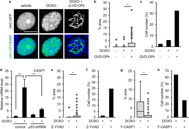
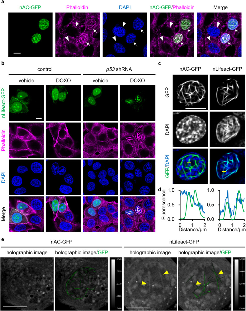
Tumor suppressor p53 plays a central role in response to DNA damage. DNA-damaging agents modulate nuclear actin dynamics, influencing cell behaviors; however, whether p53 affects the formation of nuclear actin filaments remains unclear. In this study, we found that p53 depletion promoted the formation of nuclear actin filaments in response to DNA-damaging agents, such as doxorubicin (DOXO) and etoposide (VP16). Even though the genetic probes used for the detection of nuclear actin filaments exerted a promotive effect on actin polymerization, the detected formation of nuclear actin filaments was highly dependent on both p53 depletion and DNA damage. Whilst active p53 is known to promote caspase-1 expression, the overexpression of caspase-1 reduced DNA damage-induced formation of nuclear actin filaments in p53-depleted cells. In contrast, co-treatment with DOXO and the pan-caspase inhibitor Q-VD-OPh or the caspase-1 inhibitor Z-YVAD-FMK induced the formation of nuclear actin filament formation even in cells bearing wild-type p53. These results suggest that the p53-caspase-1 axis suppresses DNA damage-induced formation of nuclear actin filaments. In addition, we found that the expression of nLifeact-GFP, the filamentous-actin-binding peptide Lifeact fused with the nuclear localization signal (NLS) and GFP, modulated the structure of nuclear actin filaments to be phalloidin-stainable in p53-depleted cells treated with the DNA-damaging agent, altering the chromatin structure and reducing the transcriptional activity. The level of phosphorylated H2AX (γH2AX), a marker of DNA damage, in these cells also reduced upon nLifeact-GFP expression, whilst details of the functional relationship between the formation of nLifeact-GFP-decorated nuclear actin filaments and DNA repair remained to be elucidated. Considering that the loss of p53 is associated with cancer progression, the results of this study raise a possibility that the artificial reinforcement of nuclear actin filaments by nLifeact-GFP may enhance the cytotoxic effect of DNA-damaging agents in aggressive cancer cells through a reduction in gene transcription.
抑癌蛋白 p53 在应对 DNA 损伤中起着核心作用。DNA 损伤试剂可调节核肌动蛋白动力学,影响细胞行为;然而,p53 是否影响核肌动蛋白丝的形成尚不清楚。在这项研究中,我们发现 p53 耗竭促进了 DNA 损伤试剂(如阿霉素(DOXO)和依托泊苷(VP16))诱导的核肌动蛋白丝的形成。尽管用于检测核肌动蛋白丝的遗传探针对肌动蛋白聚合有促进作用,但检测到的核肌动蛋白丝的形成高度依赖于 p53 耗竭和 DNA 损伤。虽然已知活性 p53 可促进半胱天冬酶-1 的表达,但 caspase-1 的过表达可降低 p53 耗竭细胞中 DNA 损伤诱导的核肌动蛋白丝的形成。相反,在 DOXO 与泛半胱天冬酶抑制剂 Q-VD-OPh 或 caspase-1 抑制剂 Z-YVAD-FMK 联合处理时,即使在携带野生型 p53 的细胞中,也会诱导核肌动蛋白丝的形成。这些结果表明,p53-caspase-1 轴抑制 DNA 损伤诱导的核肌动蛋白丝的形成。此外,我们发现,表达 nLifeact-GFP,丝状肌动蛋白结合肽 Lifeact 融合核定位信号(NLS)和 GFP,可调节核肌动蛋白丝的结构,使其在 DNA 损伤试剂处理的 p53 耗竭细胞中对鬼笔环肽染色,改变染色质结构并降低转录活性。这些细胞中磷酸化 H2AX(γH2AX)的水平(DNA 损伤的标志物)也随着 nLifeact-GFP 的表达而降低,而 nLifeact-GFP 修饰的核肌动蛋白丝形成与 DNA 修复之间的功能关系的细节仍有待阐明。考虑到 p53 的缺失与癌症进展有关,本研究的结果提出了一种可能性,即通过降低基因转录,nLifeact-GFP 增强的核肌动蛋白丝可能通过增强 DNA 损伤试剂在侵袭性癌细胞中的细胞毒性作用。