• 文献检索
  • 文档翻译
  • 深度研究
  • 学术资讯
  • Suppr Zotero 插件Zotero 插件
  • 邀请有礼
  • 套餐&价格
  • 历史记录
应用&插件
Suppr Zotero 插件Zotero 插件浏览器插件Mac 客户端Windows 客户端微信小程序
定价
高级版会员购买积分包购买API积分包
服务
文献检索文档翻译深度研究API 文档MCP 服务
关于我们
关于 Suppr公司介绍联系我们用户协议隐私条款
关注我们

Suppr 超能文献

核心技术专利:CN118964589B侵权必究
粤ICP备2023148730 号-1Suppr @ 2026

文献检索

告别复杂PubMed语法,用中文像聊天一样搜索,搜遍4000万医学文献。AI智能推荐,让科研检索更轻松。

立即免费搜索

文件翻译

保留排版,准确专业,支持PDF/Word/PPT等文件格式,支持 12+语言互译。

免费翻译文档

深度研究

AI帮你快速写综述,25分钟生成高质量综述,智能提取关键信息,辅助科研写作。

立即免费体验

抗VEGF治疗的糖尿病性黄斑水肿患者的地形图血管密度与视网膜厚度变化之间的相关性:它是一种合适的光学相干断层扫描血管造影生物标志物吗?

Correlation between Topographic Vessel Density and Retinal Thickness Changes in Patients with Diabetic Macular Edema Treated with Anti-VEGF Therapy: Is It a Suitable OCTA Biomarker?

作者信息

Santamaría Juan, Caminal José María, Cobos Estefanía, Biarnes Marc, Rodriguez-Leor Ramon, Morwani Rahul, García-Mendieta Manel, Lorenzo Daniel, García-Bru Pere, Arias Luis

机构信息

Department of Ophthalmology, Ocular Oncology and Vitreoretinal Service, Bellvitge University Hospital, L'Hospitalet de Llobregat, 08907 Barcelona, Spain.

OMIQ Research, c/Tamarit 39, 08205 Sabadell, Spain.

出版信息

J Pers Med. 2023 Dec 16;13(12):1718. doi: 10.3390/jpm13121718.

DOI:10.3390/jpm13121718
PMID:38138945
原文链接:https://pmc.ncbi.nlm.nih.gov/articles/PMC10744775/
Abstract

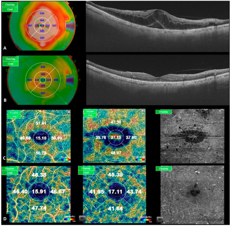
The objective of this study was to determine the correlation between topographic vessel density (VD) and retinal thickness (RT) reductions induced by vascular endothelial growth factor inhibitors (anti-VEGF) in patients with diabetic macular edema (DME) using optical coherence tomography angiography (OCTA). This was a prospective, interventional case series. VD and RT measurements were separately taken in four parafoveal subfields at baseline and after six months of treatment. This correlation was statistically assessed using Spearman's rho correlation coefficient after adjustment for multiple comparisons. The study included a total of 48 eyes in the final analysis. Mean VD decreased from baseline to month 6 (from 45.2 (±3.5) to 44.6% (±3.2) in the superficial capillary plexus and from 50 (±3.3) to 49% (±3.9) in the deep capillary plexus). Statistically significant reductions in RT were observed in all ETDRS sectors ( < 0.0001). No significant association was found between RT and VD, even when analyzing responders and non-responders separately. After six months of anti-VEGF treatment, no significant correlation was observed between the topographic VD and RT values. These findings suggest that reductions in VD values may not solely result from a reduction in microaneurysms, also being affected by the repositioning of displaced vessels due to edema and a reduction in their caliber. Therefore, VD changes may not be a suitable indirect OCTA biomarker of microaneurysm turnover and treatment response.

摘要

本研究的目的是使用光学相干断层扫描血管造影(OCTA)确定糖尿病性黄斑水肿(DME)患者中血管内皮生长因子抑制剂(抗VEGF)诱导的地形图血管密度(VD)与视网膜厚度(RT)降低之间的相关性。这是一项前瞻性干预性病例系列研究。在基线时和治疗六个月后,分别在四个黄斑旁子区域进行VD和RT测量。在对多重比较进行调整后,使用Spearman等级相关系数对这种相关性进行统计学评估。最终分析共纳入48只眼。平均VD从基线下降到第6个月(浅层毛细血管丛从45.2(±3.5)降至44.6%(±3.2),深层毛细血管丛从50(±3.3)降至49%(±3.9))。在所有ETDRS扇形区域均观察到RT有统计学意义的降低(<0.0001)。即使分别分析反应者和无反应者,也未发现RT与VD之间存在显著关联。抗VEGF治疗六个月后,地形图VD与RT值之间未观察到显著相关性。这些发现表明,VD值的降低可能不仅仅是由于微动脉瘤减少所致,还受到因水肿导致的移位血管重新定位及其管径减小的影响。因此,VD变化可能不是微动脉瘤周转率和治疗反应的合适间接OCTA生物标志物。

https://cdn.ncbi.nlm.nih.gov/pmc/blobs/6649/10744775/c1eb18915e11/jpm-13-01718-g002.jpg
https://cdn.ncbi.nlm.nih.gov/pmc/blobs/6649/10744775/680fb5369777/jpm-13-01718-g001.jpg
https://cdn.ncbi.nlm.nih.gov/pmc/blobs/6649/10744775/c1eb18915e11/jpm-13-01718-g002.jpg
https://cdn.ncbi.nlm.nih.gov/pmc/blobs/6649/10744775/680fb5369777/jpm-13-01718-g001.jpg
https://cdn.ncbi.nlm.nih.gov/pmc/blobs/6649/10744775/c1eb18915e11/jpm-13-01718-g002.jpg

相似文献

1
Correlation between Topographic Vessel Density and Retinal Thickness Changes in Patients with Diabetic Macular Edema Treated with Anti-VEGF Therapy: Is It a Suitable OCTA Biomarker?抗VEGF治疗的糖尿病性黄斑水肿患者的地形图血管密度与视网膜厚度变化之间的相关性:它是一种合适的光学相干断层扫描血管造影生物标志物吗?
J Pers Med. 2023 Dec 16;13(12):1718. doi: 10.3390/jpm13121718.
2
Changes in vessel density patterns assessed with OCTA in patients with diabetic macular edema treated with anti-VEGF therapy.接受抗血管内皮生长因子治疗的糖尿病黄斑水肿患者的 OCTA 评估血管密度模式的变化。
Acta Diabetol. 2024 Nov;61(11):1385-1392. doi: 10.1007/s00592-024-02290-5. Epub 2024 May 27.
3
Optical coherence tomography angiography analysis of macular vessel density before and after anti-VEGF therapy in eyes with diabetic retinopathy.糖尿病视网膜病变患者抗VEGF治疗前后黄斑血管密度的光学相干断层扫描血管造影分析
Int Ophthalmol. 2019 Oct;39(10):2361-2371. doi: 10.1007/s10792-019-01076-x. Epub 2019 May 22.
4
Macular vessels density in diabetic retinopathy: quantitative assessment using optical coherence tomography angiography.糖尿病视网膜病变中的黄斑血管密度:使用光学相干断层扫描血管造影术进行定量评估。
Int Ophthalmol. 2019 Aug;39(8):1845-1859. doi: 10.1007/s10792-018-1013-0. Epub 2018 Sep 7.
5
Vessel Density Changes on Optical Coherence Tomography Angiography after Vascular Endothelial Growth Factor Inhibitor Treatment for Diabetic Macular Edema.血管内皮生长因子抑制剂治疗糖尿病黄斑水肿后光学相干断层扫描血管造影的血管密度变化。
Turk J Ophthalmol. 2020 Dec 29;50(6):343-350. doi: 10.4274/tjo.galenos.2020.81592.
6
Effect of a Fixed Combination of Curcumin, Artemisia, Bromelain, and Black Pepper Oral Administration on Optical Coherence Tomography Angiography Indices in Patients with Diabetic Macular Edema.姜黄素、青蒿、菠萝蛋白酶和黑胡椒固定组合口服对糖尿病黄斑水肿患者光学相干断层扫描血管造影指数的影响。
Nutrients. 2022 Apr 6;14(7):1520. doi: 10.3390/nu14071520.
7
Optical coherence tomography angiography analysis of microvascular abnormalities and vessel density in treatment-naïve eyes with diabetic macular edema.未经治疗的糖尿病黄斑水肿患者的微血管异常和血管密度的光相干断层扫描血管造影分析。
BMC Ophthalmol. 2022 Nov 3;22(1):418. doi: 10.1186/s12886-022-02632-3.
8
Optical Coherence Tomography Angiography of DME and Its Association with Anti-VEGF Treatment Response.糖尿病性黄斑水肿的光学相干断层扫描血管造影及其与抗血管内皮生长因子治疗反应的关系。
Ophthalmology. 2016 Nov;123(11):2368-2375. doi: 10.1016/j.ophtha.2016.07.010. Epub 2016 Sep 6.
9
Optical Coherence Tomography Angiography Analysis of the Foveal Avascular Zone and Macular Vessel Density After Anti-VEGF Therapy in Eyes With Diabetic Macular Edema and Retinal Vein Occlusion.糖尿病性黄斑水肿和视网膜静脉阻塞患者抗VEGF治疗后黄斑无血管区和黄斑血管密度的光学相干断层扫描血管造影分析
Invest Ophthalmol Vis Sci. 2017 Jan 1;58(1):30-34. doi: 10.1167/iovs.16-20579.
10
Evaluation of macular vascular density and foveal avascular zone changes by optical coherence tomography angiography (OCT-A) after intravitreal dexamethasone implant in diabetic macular edema resistant to Anti-VEGF treatment.评价玻璃体内注射地塞米松植入物治疗抗血管内皮生长因子治疗抵抗的糖尿病性黄斑水肿后的黄斑血管密度和中心无血管区变化的光学相干断层扫描血管造影(OCT-A)。
Int Ophthalmol. 2022 Nov;42(11):3579-3588. doi: 10.1007/s10792-022-02374-7. Epub 2022 Jun 23.

引用本文的文献

1
Differential artery-vein analysis in OCTA for predicting the anti-VEGF treatment outcome of diabetic macular edema.光学相干断层扫描血管造影中动脉-静脉差异分析预测糖尿病性黄斑水肿抗血管内皮生长因子治疗效果
Biomed Opt Express. 2025 Apr 1;16(4):1732-1741. doi: 10.1364/BOE.557748.
2
The macular microvascular changes in children with a history of retinopathy of prematurity treated with intravitreal Bevacizumab injection using optical coherence tomography angiography: a retrospective cohort.经玻璃体腔注射贝伐单抗治疗早产儿视网膜病变后儿童的黄斑微血管改变:回顾性队列研究。
BMC Ophthalmol. 2024 Nov 19;24(1):505. doi: 10.1186/s12886-024-03773-3.

本文引用的文献

1
Anti-VEGF and steroid combination therapy relative to anti-VEGF mono therapy for the treatment of refractory DME: A systematic review of efficacy and meta-analysis of safety.抗 VEGF 和类固醇联合治疗与抗 VEGF 单药治疗难治性 DME 的疗效比较:系统评价和安全性的荟萃分析。
Acta Ophthalmol. 2024 May;102(3):e204-e214. doi: 10.1111/aos.15724. Epub 2023 Jun 26.
2
Role of Microaneurysms in the Pathogenesis and Therapy of Diabetic Macular Edema: A Descriptive Review.微动脉瘤在糖尿病性黄斑水肿发病机制和治疗中的作用:描述性综述。
Medicina (Kaunas). 2023 Feb 22;59(3):435. doi: 10.3390/medicina59030435.
3
Prognostic factors for the development and progression of proliferative diabetic retinopathy in people with diabetic retinopathy.
增生性糖尿病性视网膜病变在糖尿病性视网膜病变患者中发展和进展的预测因素。
Cochrane Database Syst Rev. 2023 Feb 22;2(2):CD013775. doi: 10.1002/14651858.CD013775.pub2.
4
Evaluation of microaneurysms as predictors of therapeutic response to anti-VEGF therapy in patients with DME.评估微动脉瘤作为 DME 患者抗 VEGF 治疗反应的预测因子。
PLoS One. 2022 Nov 28;17(11):e0277920. doi: 10.1371/journal.pone.0277920. eCollection 2022.
5
Regional Variety of Reduction in Retinal Thickness of Diabetic Macular Edema after Anti-VEGF Treatment.抗血管内皮生长因子治疗后糖尿病黄斑水肿视网膜厚度降低的区域性差异。
Medicina (Kaunas). 2022 Jul 14;58(7):933. doi: 10.3390/medicina58070933.
6
OCTA changes following loading phase with intravitreal aflibercept for DME.OCTA 检查在接受玻璃体内注射阿柏西普治疗 DME 的负荷期后的改变。
Can J Ophthalmol. 2023 Oct;58(5):480-490. doi: 10.1016/j.jcjo.2022.04.005. Epub 2022 May 5.
7
Treatment of Diabetic Macular Edema with Aflibercept and Micropulse Laser (DAM Study).阿柏西普与微脉冲激光治疗糖尿病性黄斑水肿(DAM研究)
Clin Ophthalmol. 2022 Apr 8;16:1109-1115. doi: 10.2147/OPTH.S360869. eCollection 2022.
8
Microaneurysm Turnover in Mild Non-Proliferative Diabetic Retinopathy is Associated with Progression and Development of Vision-Threatening Complications: A 5-Year Longitudinal Study.轻度非增殖性糖尿病视网膜病变中微动脉瘤的周转率与视力威胁性并发症的进展和发生相关:一项5年纵向研究
J Clin Med. 2021 May 15;10(10):2142. doi: 10.3390/jcm10102142.
9
Optical Coherence Tomography (Angiography) Biomarkers in the Assessment and Monitoring of Diabetic Macular Edema.光学相干断层扫描(血管造影)生物标志物在糖尿病性黄斑水肿评估和监测中的应用。
J Diabetes Res. 2020 Dec 31;2020:6655021. doi: 10.1155/2020/6655021. eCollection 2020.
10
Vessel Density Changes on Optical Coherence Tomography Angiography after Vascular Endothelial Growth Factor Inhibitor Treatment for Diabetic Macular Edema.血管内皮生长因子抑制剂治疗糖尿病黄斑水肿后光学相干断层扫描血管造影的血管密度变化。
Turk J Ophthalmol. 2020 Dec 29;50(6):343-350. doi: 10.4274/tjo.galenos.2020.81592.