• 文献检索
  • 文档翻译
  • 深度研究
  • 学术资讯
  • Suppr Zotero 插件Zotero 插件
  • 邀请有礼
  • 套餐&价格
  • 历史记录
应用&插件
Suppr Zotero 插件Zotero 插件浏览器插件Mac 客户端Windows 客户端微信小程序
定价
高级版会员购买积分包购买API积分包
服务
文献检索文档翻译深度研究API 文档MCP 服务
关于我们
关于 Suppr公司介绍联系我们用户协议隐私条款
关注我们

Suppr 超能文献

核心技术专利:CN118964589B侵权必究
粤ICP备2023148730 号-1Suppr @ 2026

文献检索

告别复杂PubMed语法,用中文像聊天一样搜索,搜遍4000万医学文献。AI智能推荐,让科研检索更轻松。

立即免费搜索

文件翻译

保留排版,准确专业,支持PDF/Word/PPT等文件格式,支持 12+语言互译。

免费翻译文档

深度研究

AI帮你快速写综述,25分钟生成高质量综述,智能提取关键信息,辅助科研写作。

立即免费体验

基于机器学习的放射组学分析和生长可视化在随访 CT 中用于消融部位复发的诊断。

Machine learning-based radiomic analysis and growth visualization for ablation site recurrence diagnosis in follow-up CT.

机构信息

Department of Radiology, Medical Imaging Center Groningen, University of Groningen, University Medical Center Groningen, PO Box 30001, 9700 RB, Groningen, The Netherlands.

Diagnostic Image Analysis Group, Department of Medical Imaging, Radboud University Medical Center, 6500 HB, Nijmegen, The Netherlands.

出版信息

Abdom Radiol (NY). 2024 Apr;49(4):1122-1131. doi: 10.1007/s00261-023-04178-4. Epub 2024 Jan 30.

DOI:10.1007/s00261-023-04178-4
PMID:38289352
原文链接:https://pmc.ncbi.nlm.nih.gov/articles/PMC10955006/
Abstract

OBJECTIVES

Detecting ablation site recurrence (ASR) after thermal ablation remains a challenge for radiologists due to the similarity between tumor recurrence and post-ablative changes. Radiomic analysis and machine learning methods may show additional value in addressing this challenge. The present study primarily sought to determine the efficacy of radiomic analysis in detecting ASR on follow-up computed tomography (CT) scans. The second aim was to develop a visualization tool capable of emphasizing regions of ASR between follow-up scans in individual patients.

MATERIALS AND METHODS

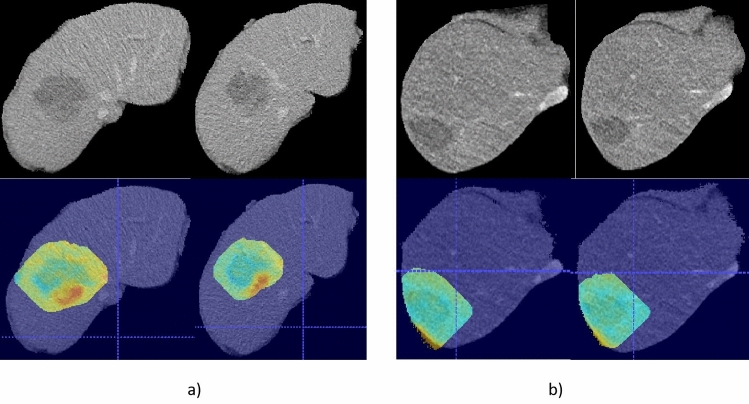
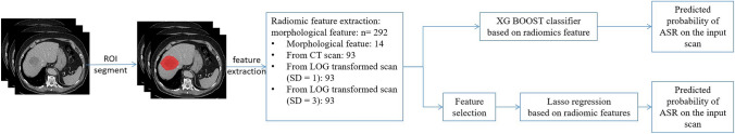
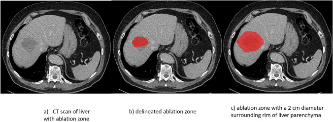
Lasso regression and Extreme Gradient Boosting (XGBoost) classifiers were employed for modeling radiomic features extracted from regions of interest delineated by two radiologists. A leave-one-out test (LOOT) was utilized for performance evaluation. A visualization method, creating difference heatmaps (diff-maps) between two follow-up scans, was developed to emphasize regions of growth and thereby highlighting potential ASR.

RESULTS

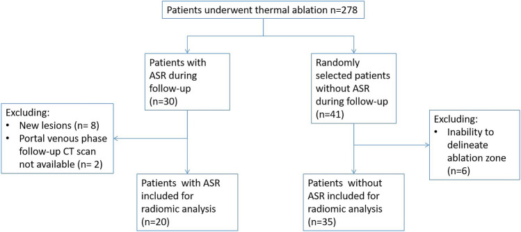
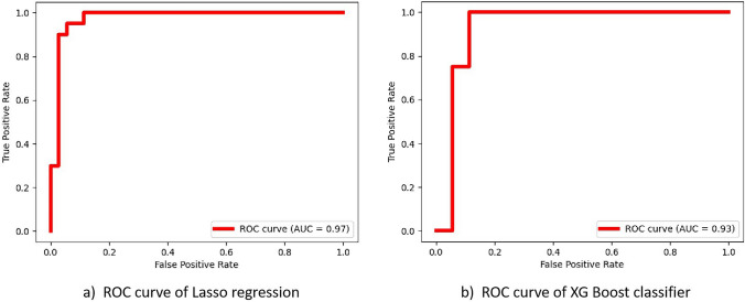
A total of 55 patients, including 20 with and 35 without ASR, were included in the radiomic analysis. The best performing model was achieved by Lasso regression tested with the LOOT approach, reaching an area under the curve (AUC) of 0.97 and an accuracy of 92.73%. The XGBoost classifier demonstrated better performance when trained with all extracted radiomic features than without feature selection, achieving an AUC of 0.93 and an accuracy of 89.09%. The diff-maps correctly highlighted post-ablative liver tumor recurrence in all patients.

CONCLUSIONS

Machine learning-based radiomic analysis and growth visualization proved effective in detecting ablation site recurrence on follow-up CT scans.

摘要

目的

由于肿瘤复发与消融后改变之间存在相似性,热消融后消融部位复发(ASR)的检测对放射科医生来说仍是一项挑战。放射组学分析和机器学习方法可能在解决这一挑战方面具有额外的价值。本研究主要旨在确定放射组学分析在检测后续 CT 扫描中 ASR 的功效。第二个目的是开发一种可视化工具,能够在个体患者的随访扫描中突出显示 ASR 区域。

材料与方法

使用套索回归和极端梯度提升(XGBoost)分类器对由两位放射科医生勾画的感兴趣区域的放射组学特征进行建模。采用留一法(LOOT)进行性能评估。开发了一种创建两次随访扫描之间差异热图(diff-maps)的可视化方法,以强调生长区域,从而突出潜在的 ASR。

结果

共纳入 55 例患者,其中 20 例有 ASR,35 例无 ASR。对 Lasso 回归进行 LOOT 测试的最佳模型,曲线下面积(AUC)为 0.97,准确率为 92.73%。XGBoost 分类器在使用所有提取的放射组学特征进行训练时的性能优于不进行特征选择,AUC 为 0.93,准确率为 89.09%。diff-maps 正确地突出了所有患者消融后肝肿瘤的复发。

结论

基于机器学习的放射组学分析和生长可视化在检测后续 CT 扫描中的消融部位复发方面是有效的。

https://cdn.ncbi.nlm.nih.gov/pmc/blobs/125d/10955006/4a0af3caf57d/261_2023_4178_Fig5_HTML.jpg
https://cdn.ncbi.nlm.nih.gov/pmc/blobs/125d/10955006/20593e6c2fca/261_2023_4178_Fig1_HTML.jpg
https://cdn.ncbi.nlm.nih.gov/pmc/blobs/125d/10955006/2712bd8b7545/261_2023_4178_Fig2_HTML.jpg
https://cdn.ncbi.nlm.nih.gov/pmc/blobs/125d/10955006/f1181b04458c/261_2023_4178_Fig3_HTML.jpg
https://cdn.ncbi.nlm.nih.gov/pmc/blobs/125d/10955006/470c47661bce/261_2023_4178_Fig4_HTML.jpg
https://cdn.ncbi.nlm.nih.gov/pmc/blobs/125d/10955006/4a0af3caf57d/261_2023_4178_Fig5_HTML.jpg

相似文献

1
Machine learning-based radiomic analysis and growth visualization for ablation site recurrence diagnosis in follow-up CT.基于机器学习的放射组学分析和生长可视化在随访 CT 中用于消融部位复发的诊断。
Abdom Radiol (NY). 2024 Apr;49(4):1122-1131. doi: 10.1007/s00261-023-04178-4. Epub 2024 Jan 30.
2
Radiomic analysis for early differentiation of lung cancer recurrence from fibrosis in patients treated with lung stereotactic ablative radiotherapy.基于放射组学分析的肺立体定向消融放疗后患者肺纤维化与肺癌复发的早期鉴别
Phys Med Biol. 2023 Aug 7;68(16). doi: 10.1088/1361-6560/acd431.
3
Develop a radiomics-based machine learning model to predict the stone-free rate post-percutaneous nephrolithotomy.建立基于放射组学的机器学习模型,以预测经皮肾镜取石术后的无石率。
Urolithiasis. 2024 Apr 13;52(1):64. doi: 10.1007/s00240-024-01562-7.
4
Distinguishing granulomas from adenocarcinomas by integrating stable and discriminating radiomic features on non-contrast computed tomography scans.基于非增强 CT 扫描的稳定且具有鉴别能力的放射组学特征区分肉芽肿与腺癌。
Eur J Cancer. 2021 May;148:146-158. doi: 10.1016/j.ejca.2021.02.008. Epub 2021 Mar 17.
5
The impact of the combat method on radiomics feature compensation and analysis of scanners from different manufacturers.战斗方法对不同制造商扫描仪的放射组学特征补偿和分析的影响。
BMC Med Imaging. 2024 Jun 6;24(1):137. doi: 10.1186/s12880-024-01306-4.
6
Optimizing the radiomics-machine-learning model based on non-contrast enhanced CT for the simplified risk categorization of thymic epithelial tumors: A large cohort retrospective study.基于非增强CT优化放射组学-机器学习模型用于胸腺瘤简化风险分类:一项大型队列回顾性研究
Lung Cancer. 2022 Apr;166:150-160. doi: 10.1016/j.lungcan.2022.03.007. Epub 2022 Mar 8.
7
Optimal radiological gallbladder lesion characterization by combining visual assessment with CT-based radiomics.联合视觉评估与 CT 影像组学对胆囊病变进行最佳影像学特征描述。
Eur Radiol. 2023 Apr;33(4):2725-2734. doi: 10.1007/s00330-022-09281-6. Epub 2022 Nov 25.
8
Application of machine learning model to predict osteoporosis based on abdominal computed tomography images of the psoas muscle: a retrospective study.基于腰部竖脊肌 CT 图像的机器学习模型预测骨质疏松症的应用:一项回顾性研究。
BMC Geriatr. 2022 Oct 13;22(1):796. doi: 10.1186/s12877-022-03502-9.
9
Machine learning in the differentiation of follicular lymphoma from diffuse large B-cell lymphoma with radiomic [F]FDG PET/CT features.基于放射组学[F]FDG PET/CT特征的机器学习在滤泡性淋巴瘤与弥漫性大B细胞淋巴瘤鉴别诊断中的应用
Eur J Nucl Med Mol Imaging. 2022 Apr;49(5):1535-1543. doi: 10.1007/s00259-021-05626-3. Epub 2021 Dec 1.
10
Machine-learning-based computed tomography radiomic analysis for histologic subtype classification of thymic epithelial tumours.基于机器学习的 CT 影像组学分析用于胸腺癌组织学分型。
Eur J Radiol. 2020 May;126:108929. doi: 10.1016/j.ejrad.2020.108929. Epub 2020 Mar 2.

引用本文的文献

1
Applications of artificial intelligence in interventional oncology: An up-to-date review of the literature.人工智能在介入肿瘤学中的应用:文献综述
Jpn J Radiol. 2025 Feb;43(2):164-176. doi: 10.1007/s11604-024-01668-3. Epub 2024 Oct 2.

本文引用的文献

1
A prospective multicentre trial on survival after Microwave Ablation VErsus Resection for Resectable Colorectal liver metastases (MAVERRIC).微波消融与切除术治疗可切除结直肠癌肝转移的生存前瞻性多中心研究(MAVERRIC)。
Eur J Cancer. 2023 Jul;187:65-76. doi: 10.1016/j.ejca.2023.03.038. Epub 2023 Apr 5.
2
Introduction to radiomics for a clinical audience.放射组学简介:面向临床受众
Clin Radiol. 2023 Feb;78(2):83-98. doi: 10.1016/j.crad.2022.08.149.
3
Ablation versus resection for resectable colorectal liver metastases - Health care related cost and survival analyses from a quasi-randomised study.
可切除结直肠癌肝转移的消融与切除——一项准随机研究的医疗相关成本和生存分析
Eur J Surg Oncol. 2023 Feb;49(2):416-425. doi: 10.1016/j.ejso.2022.09.006. Epub 2022 Sep 13.
4
Recurrent Hepatocellular Carcinoma: Patterns, Detection, Staging and Treatment.复发性肝细胞癌:模式、检测、分期与治疗
J Hepatocell Carcinoma. 2022 Sep 3;9:947-957. doi: 10.2147/JHC.S342266. eCollection 2022.
5
Hepatocellular carcinoma.肝细胞癌
Lancet. 2022 Oct 15;400(10360):1345-1362. doi: 10.1016/S0140-6736(22)01200-4. Epub 2022 Sep 6.
6
CT-Based Radiomics for the Recurrence Prediction of Hepatocellular Carcinoma After Surgical Resection.基于CT的放射组学用于预测肝细胞癌手术切除后的复发
J Hepatocell Carcinoma. 2022 May 23;9:453-465. doi: 10.2147/JHC.S362772. eCollection 2022.
7
Colorectal liver metastasis: molecular mechanism and interventional therapy.结直肠癌肝转移:分子机制与介入治疗。
Signal Transduct Target Ther. 2022 Mar 4;7(1):70. doi: 10.1038/s41392-022-00922-2.
8
Outcomes after primary and repeat thermal ablation of hepatocellular carcinoma with or without liver transplantation.肝细胞癌初次及重复热消融联合或不联合肝移植后的结局
Eur Radiol. 2022 Jun;32(6):4168-4176. doi: 10.1007/s00330-021-08515-3. Epub 2022 Feb 8.
9
3D Quantitative Ablation Margins for Prediction of Ablation Site Recurrence After Stereotactic Image-Guided Microwave Ablation of Colorectal Liver Metastases: A Multicenter Study.3D定量消融边缘用于预测立体定向图像引导下微波消融结直肠癌肝转移灶后消融部位复发:一项多中心研究
Front Oncol. 2021 Nov 15;11:757167. doi: 10.3389/fonc.2021.757167. eCollection 2021.
10
Stereotactic and Robotic Minimally Invasive Thermal Ablation of Malignant Liver Tumors: A Systematic Review and Meta-Analysis.立体定向和机器人辅助微创热消融治疗恶性肝肿瘤:一项系统评价和荟萃分析。
Front Oncol. 2021 Sep 23;11:713685. doi: 10.3389/fonc.2021.713685. eCollection 2021.