• 文献检索
  • 文档翻译
  • 深度研究
  • 学术资讯
  • Suppr Zotero 插件Zotero 插件
  • 邀请有礼
  • 套餐&价格
  • 历史记录
应用&插件
Suppr Zotero 插件Zotero 插件浏览器插件Mac 客户端Windows 客户端微信小程序
定价
高级版会员购买积分包购买API积分包
服务
文献检索文档翻译深度研究API 文档MCP 服务
关于我们
关于 Suppr公司介绍联系我们用户协议隐私条款
关注我们

Suppr 超能文献

核心技术专利:CN118964589B侵权必究
粤ICP备2023148730 号-1Suppr @ 2026

文献检索

告别复杂PubMed语法,用中文像聊天一样搜索,搜遍4000万医学文献。AI智能推荐,让科研检索更轻松。

立即免费搜索

文件翻译

保留排版,准确专业,支持PDF/Word/PPT等文件格式,支持 12+语言互译。

免费翻译文档

深度研究

AI帮你快速写综述,25分钟生成高质量综述,智能提取关键信息,辅助科研写作。

立即免费体验

冷冻电镜结构解析揭示了人源 CLC-2 电压门控氯离子通道的球链门控机制。

CryoEM structures of the human CLC-2 voltage-gated chloride channel reveal a ball-and-chain gating mechanism.

机构信息

Department of Molecular and Cellular Physiology, Stanford University, Stanford, United States.

Department of Chemistry, Stanford University, Stanford, United States.

出版信息

Elife. 2024 Feb 12;12:RP90648. doi: 10.7554/eLife.90648.

DOI:10.7554/eLife.90648
PMID:38345841
原文链接:https://pmc.ncbi.nlm.nih.gov/articles/PMC10942593/
Abstract

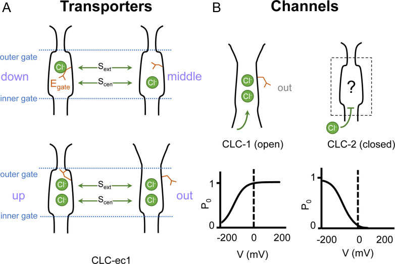
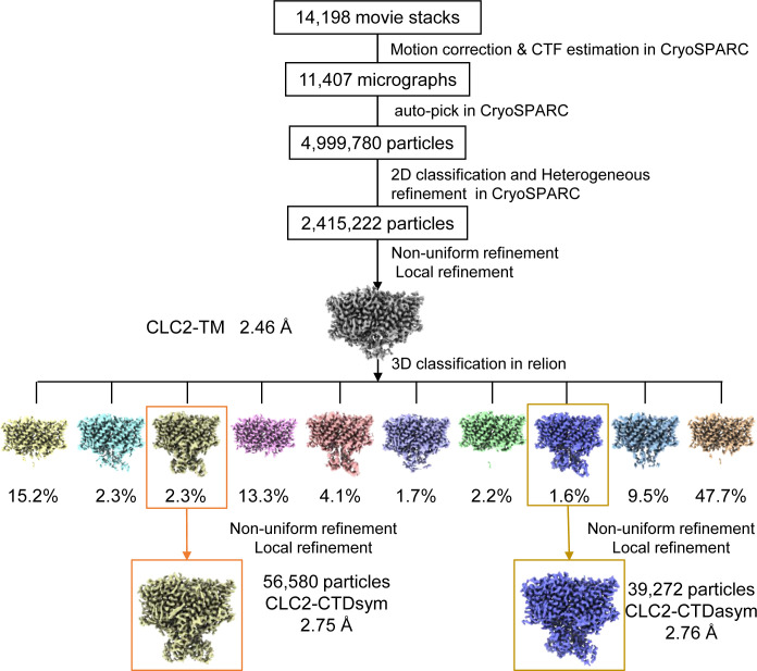
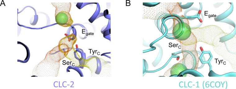
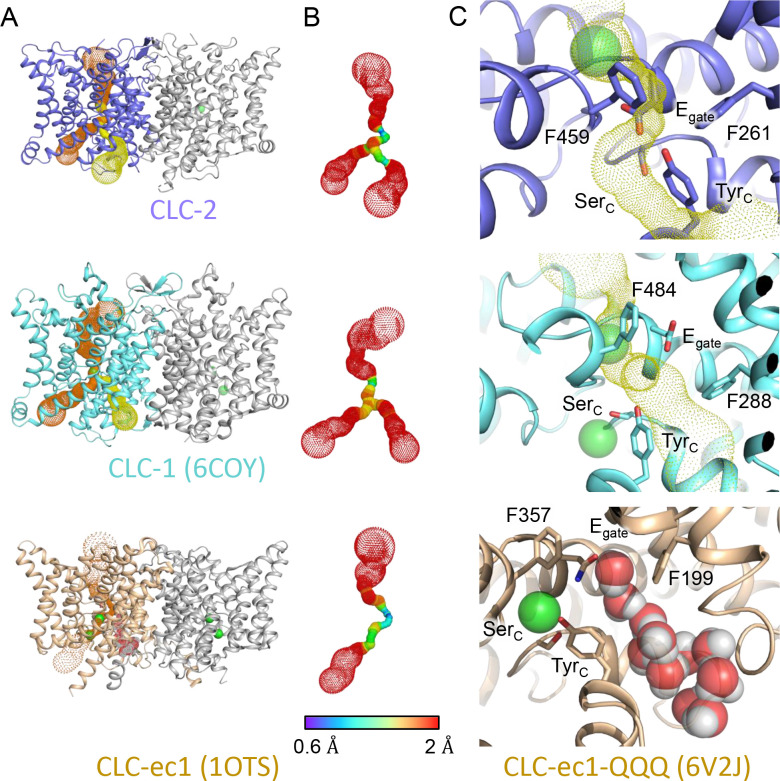
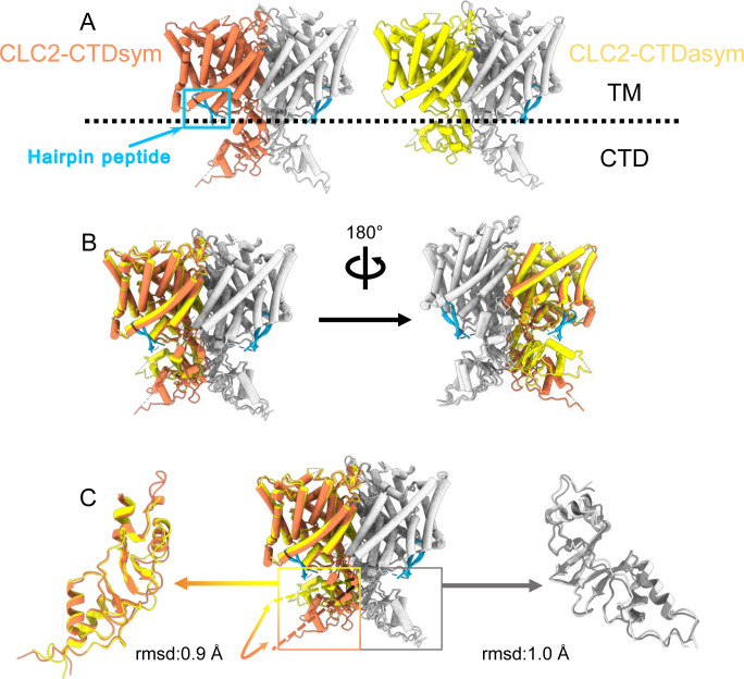
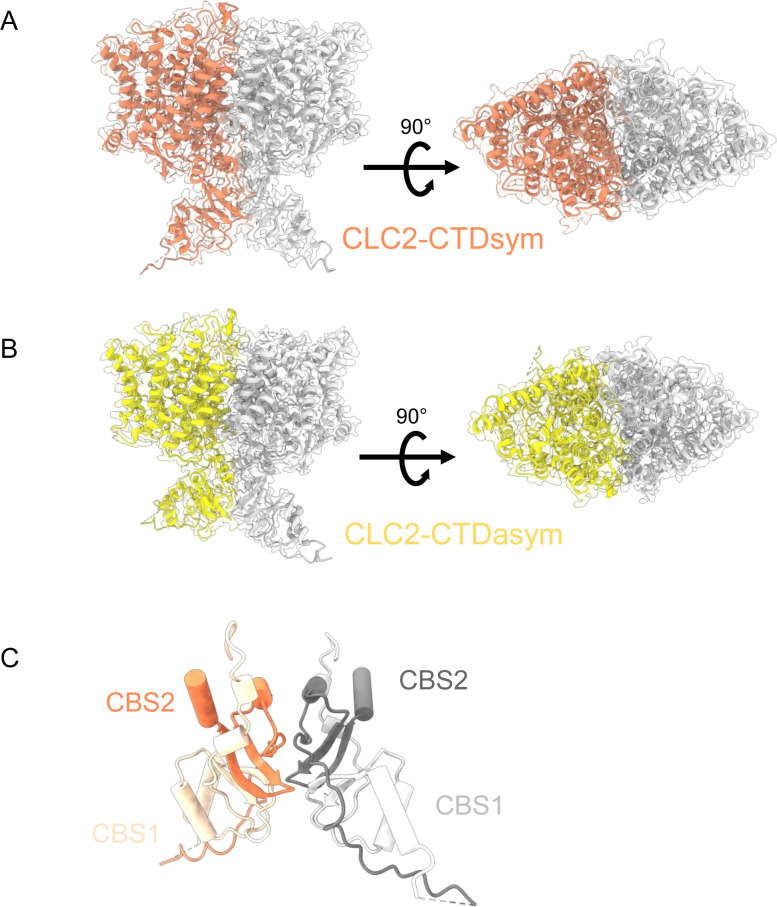
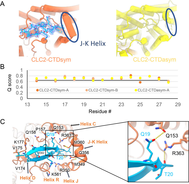
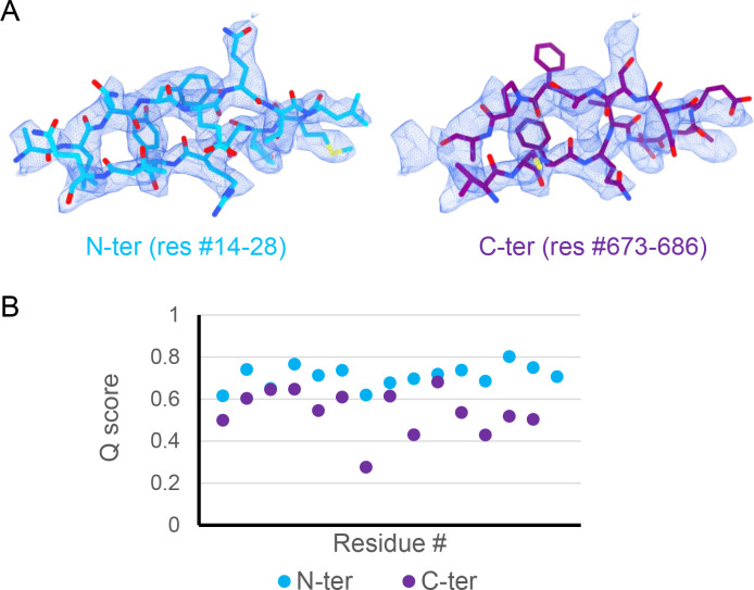
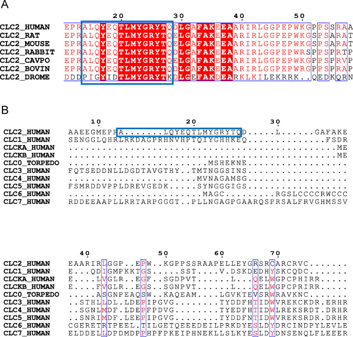
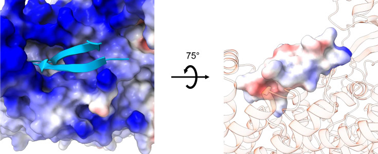
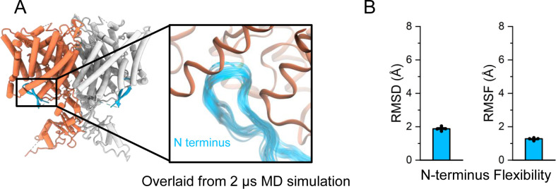
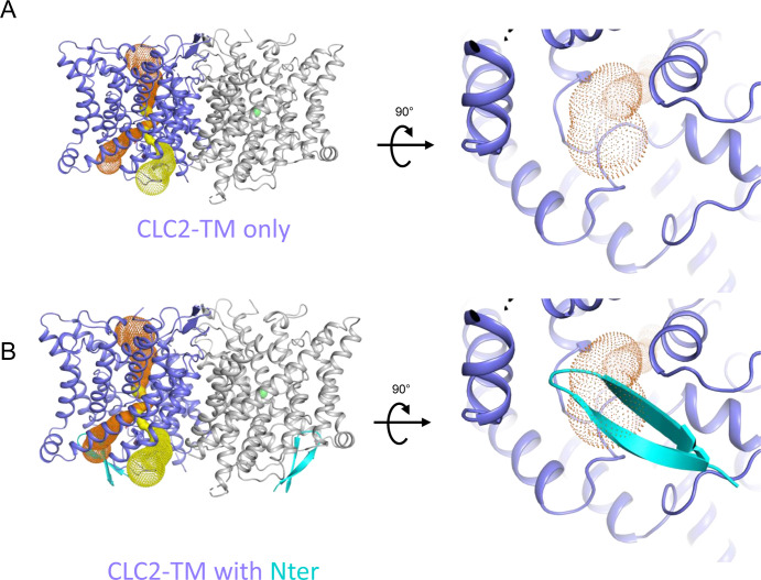
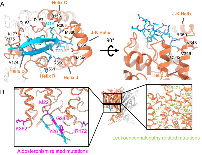
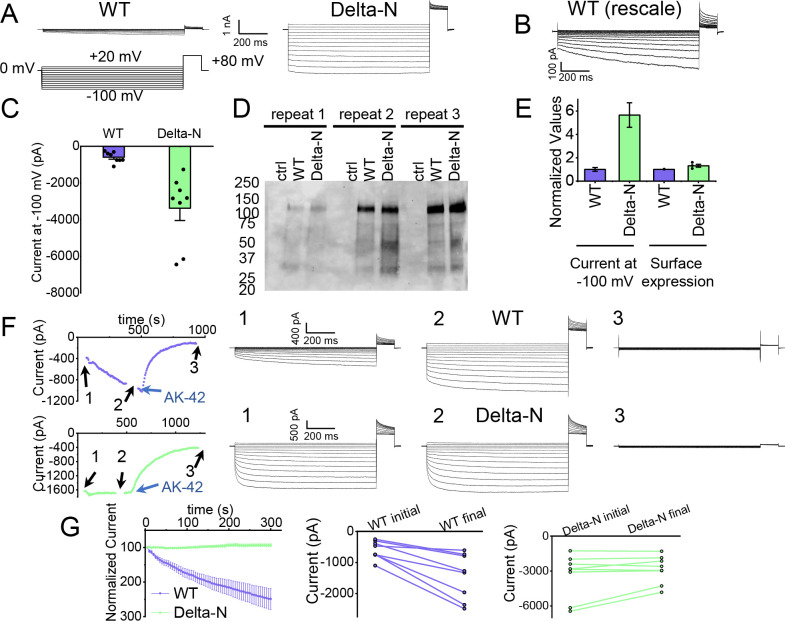
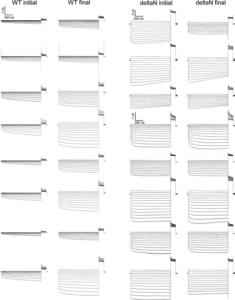
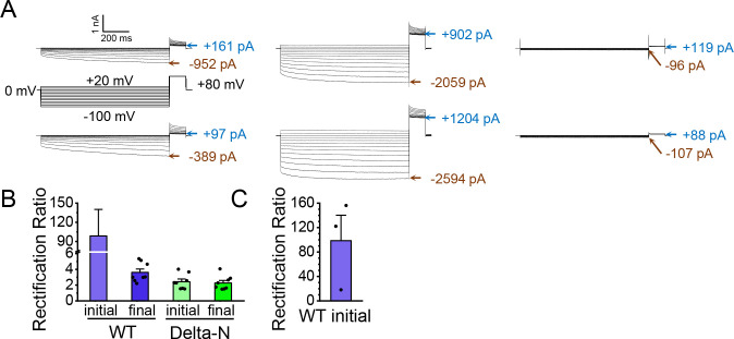
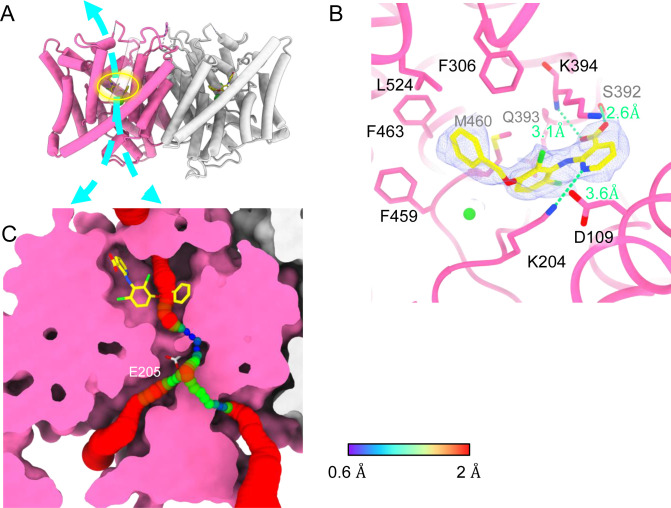
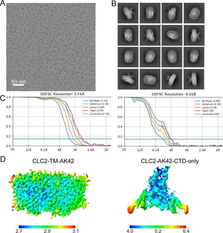
CLC-2 is a voltage-gated chloride channel that contributes to electrical excitability and ion homeostasis in many different tissues. Among the nine mammalian CLC homologs, CLC-2 is uniquely activated by hyperpolarization, rather than depolarization, of the plasma membrane. The molecular basis for the divergence in polarity of voltage gating among closely related homologs has been a long-standing mystery, in part because few CLC channel structures are available. Here, we report cryoEM structures of human CLC-2 at 2.46 - 2.76 Å, in the presence and absence of the selective inhibitor AK-42. AK-42 binds within the extracellular entryway of the Cl-permeation pathway, occupying a pocket previously proposed through computational docking studies. In the apo structure, we observed two distinct conformations involving rotation of one of the cytoplasmic C-terminal domains (CTDs). In the absence of CTD rotation, an intracellular N-terminal 15-residue hairpin peptide nestles against the TM domain to physically occlude the Cl-permeation pathway. This peptide is highly conserved among species variants of CLC-2 but is not present in other CLC homologs. Previous studies suggested that the N-terminal domain of CLC-2 influences channel properties via a "ball-and-chain" gating mechanism, but conflicting data cast doubt on such a mechanism, and thus the structure of the N-terminal domain and its interaction with the channel has been uncertain. Through electrophysiological studies of an N-terminal deletion mutant lacking the 15-residue hairpin peptide, we support a model in which the N-terminal hairpin of CLC-2 stabilizes a closed state of the channel by blocking the cytoplasmic Cl-permeation pathway.

摘要

CLC-2 是一种电压门控氯离子通道,它有助于许多不同组织的电兴奋性和离子内稳态。在哺乳动物的 9 种 CLC 同源物中,CLC-2 独特地被细胞膜的超极化而不是去极化激活。在密切相关的同源物中,电压门控极性差异的分子基础一直是一个长期存在的谜,部分原因是很少有 CLC 通道结构可用。在这里,我们报告了在存在和不存在选择性抑制剂 AK-42 的情况下,人 CLC-2 的 2.46-2.76Å 的冷冻电镜结构。AK-42 结合在氯离子渗透途径的细胞外入口内,占据了以前通过计算对接研究提出的口袋。在apo 结构中,我们观察到两种涉及一个细胞质 C 端结构域(CTD)旋转的不同构象。在没有 CTD 旋转的情况下,一个细胞内的 N 端 15 个残基发夹肽紧贴 TM 域,物理上阻塞了氯离子渗透途径。该肽在 CLC-2 的物种变体中高度保守,但在其他 CLC 同源物中不存在。以前的研究表明,CLC-2 的 N 端结构域通过“球链”门控机制影响通道特性,但相互矛盾的数据对这种机制提出了质疑,因此 N 端结构域及其与通道的相互作用一直不确定。通过对缺失了 15 个残基发夹肽的 N 端缺失突变体的电生理研究,我们支持这样一种模型,即 CLC-2 的 N 端发夹通过阻塞细胞质氯离子渗透途径来稳定通道的关闭状态。

https://cdn.ncbi.nlm.nih.gov/pmc/blobs/193b/10942593/0b57dc7c7387/elife-90648-fig7-figsupp4.jpg
https://cdn.ncbi.nlm.nih.gov/pmc/blobs/193b/10942593/c472149e294a/elife-90648-fig1.jpg
https://cdn.ncbi.nlm.nih.gov/pmc/blobs/193b/10942593/b839cb595827/elife-90648-fig2.jpg
https://cdn.ncbi.nlm.nih.gov/pmc/blobs/193b/10942593/5571f363f2f0/elife-90648-fig2-figsupp1.jpg
https://cdn.ncbi.nlm.nih.gov/pmc/blobs/193b/10942593/d69171d95571/elife-90648-fig2-figsupp2.jpg
https://cdn.ncbi.nlm.nih.gov/pmc/blobs/193b/10942593/4687918e13b7/elife-90648-fig2-figsupp3.jpg
https://cdn.ncbi.nlm.nih.gov/pmc/blobs/193b/10942593/f4970df2efc3/elife-90648-fig2-figsupp4.jpg
https://cdn.ncbi.nlm.nih.gov/pmc/blobs/193b/10942593/244de05e0241/elife-90648-fig3.jpg
https://cdn.ncbi.nlm.nih.gov/pmc/blobs/193b/10942593/e87e4b82f464/elife-90648-fig3-figsupp1.jpg
https://cdn.ncbi.nlm.nih.gov/pmc/blobs/193b/10942593/5929dd1a8de0/elife-90648-fig3-figsupp2.jpg
https://cdn.ncbi.nlm.nih.gov/pmc/blobs/193b/10942593/30dd58d838a9/elife-90648-fig4.jpg
https://cdn.ncbi.nlm.nih.gov/pmc/blobs/193b/10942593/295257828317/elife-90648-fig4-figsupp1.jpg
https://cdn.ncbi.nlm.nih.gov/pmc/blobs/193b/10942593/02b1bf75ba67/elife-90648-fig4-figsupp2.jpg
https://cdn.ncbi.nlm.nih.gov/pmc/blobs/193b/10942593/4189d7185181/elife-90648-fig4-figsupp3.jpg
https://cdn.ncbi.nlm.nih.gov/pmc/blobs/193b/10942593/60a0b05da8d8/elife-90648-fig4-figsupp4.jpg
https://cdn.ncbi.nlm.nih.gov/pmc/blobs/193b/10942593/74f68726f237/elife-90648-fig4-figsupp5.jpg
https://cdn.ncbi.nlm.nih.gov/pmc/blobs/193b/10942593/97eb4e98af36/elife-90648-fig5.jpg
https://cdn.ncbi.nlm.nih.gov/pmc/blobs/193b/10942593/fc1566b31dae/elife-90648-fig5-figsupp1.jpg
https://cdn.ncbi.nlm.nih.gov/pmc/blobs/193b/10942593/9caf53503536/elife-90648-fig5-figsupp2.jpg
https://cdn.ncbi.nlm.nih.gov/pmc/blobs/193b/10942593/4efbab335f72/elife-90648-fig5-figsupp3.jpg
https://cdn.ncbi.nlm.nih.gov/pmc/blobs/193b/10942593/03b0f1dba1eb/elife-90648-fig5-figsupp4.jpg
https://cdn.ncbi.nlm.nih.gov/pmc/blobs/193b/10942593/0dcb4d9b78ea/elife-90648-fig5-figsupp5.jpg
https://cdn.ncbi.nlm.nih.gov/pmc/blobs/193b/10942593/00d8208027a9/elife-90648-fig5-figsupp6.jpg
https://cdn.ncbi.nlm.nih.gov/pmc/blobs/193b/10942593/cf41506822e5/elife-90648-fig6.jpg
https://cdn.ncbi.nlm.nih.gov/pmc/blobs/193b/10942593/6d0c5b4175e6/elife-90648-fig6-figsupp1.jpg
https://cdn.ncbi.nlm.nih.gov/pmc/blobs/193b/10942593/9ea9db9e25c4/elife-90648-fig6-figsupp2.jpg
https://cdn.ncbi.nlm.nih.gov/pmc/blobs/193b/10942593/e405f72d8b89/elife-90648-fig7.jpg
https://cdn.ncbi.nlm.nih.gov/pmc/blobs/193b/10942593/b5670035cab7/elife-90648-fig7-figsupp2.jpg
https://cdn.ncbi.nlm.nih.gov/pmc/blobs/193b/10942593/0b57dc7c7387/elife-90648-fig7-figsupp4.jpg

相似文献

1
CryoEM structures of the human CLC-2 voltage-gated chloride channel reveal a ball-and-chain gating mechanism.冷冻电镜结构解析揭示了人源 CLC-2 电压门控氯离子通道的球链门控机制。
Elife. 2024 Feb 12;12:RP90648. doi: 10.7554/eLife.90648.
2
CryoEM structures of the human CLC-2 voltage gated chloride channel reveal a ball and chain gating mechanism.人类CLC-2电压门控氯离子通道的冷冻电镜结构揭示了一种球链门控机制。
bioRxiv. 2023 Nov 29:2023.08.13.553136. doi: 10.1101/2023.08.13.553136.
3
Emerging issues of connexin channels: biophysics fills the gap.连接蛋白通道的新问题:生物物理学填补空白。
Q Rev Biophys. 2001 Aug;34(3):325-472. doi: 10.1017/s0033583501003705.
4
Cryo-EM structures of ClC-2 chloride channel reveal the blocking mechanism of its specific inhibitor AK-42.冷冻电镜结构揭示 ClC-2 氯离子通道及其特异性抑制剂 AK-42 的作用机制。
Nat Commun. 2023 Jun 9;14(1):3424. doi: 10.1038/s41467-023-39218-6.
5
Chloride dependence of hyperpolarization-activated chloride channel gates.超极化激活氯离子通道门控对氯离子的依赖性
J Physiol. 1999 Mar 1;515 ( Pt 2)(Pt 2):341-53. doi: 10.1111/j.1469-7793.1999.341ac.x.
6
Effect of an N-terminus deletion on voltage-dependent gating of the ClC-2 chloride channel.N 端缺失对 ClC-2 氯离子通道电压依赖性门控的影响。
J Physiol. 2002 Oct 15;544(2):363-72. doi: 10.1113/jphysiol.2002.026096.
7
Quantitative analysis of the voltage-dependent gating of mouse parotid ClC-2 chloride channel.小鼠腮腺ClC-2氯离子通道电压依赖性门控的定量分析
J Gen Physiol. 2005 Dec;126(6):591-603. doi: 10.1085/jgp.200509310. Epub 2005 Nov 14.
8
Determinants of slow gating in ClC-0, the voltage-gated chloride channel of Torpedo marmorata.电鳐电压门控氯离子通道ClC-0慢速门控的决定因素。
Am J Physiol. 1998 Apr;274(4):C966-73. doi: 10.1152/ajpcell.1998.274.4.C966.
9
Nonequilibrium gating and voltage dependence of the ClC-0 Cl- channel.ClC-0氯离子通道的非平衡门控与电压依赖性
J Gen Physiol. 1996 Oct;108(4):237-50. doi: 10.1085/jgp.108.4.237.
10
Inward rectification in ClC-0 chloride channels caused by mutations in several protein regions.由几个蛋白质区域的突变导致的ClC-0氯离子通道内向整流。
J Gen Physiol. 1997 Aug;110(2):165-71. doi: 10.1085/jgp.110.2.165.

引用本文的文献

1
NT2-derived astrocyte-neuron co-culture reflects physiological relevance and offers research validity.源自NT2的星形胶质细胞-神经元共培养反映了生理相关性并提供了研究有效性。
Cell Mol Biol Lett. 2025 Jul 25;30(1):90. doi: 10.1186/s11658-025-00765-z.
2
Structural basis of ClC-3 transporter inhibition by TMEM9 and PtdIns(3,5)P.TMEM9和磷脂酰肌醇-3,5-二磷酸对氯离子通道蛋白3(ClC-3)转运体的抑制作用的结构基础
Nat Struct Mol Biol. 2025 Jul 16. doi: 10.1038/s41594-025-01617-2.
3
Molecular mechanism of exchange coupling in CLC chloride/proton antiporters.

本文引用的文献

1
ClC-1 Chloride Channel: Inputs on the Structure-Function Relationship of Myotonia Congenita-Causing Mutations.ClC-1氯离子通道:先天性肌强直致病突变结构-功能关系的研究进展
Biomedicines. 2023 Sep 24;11(10):2622. doi: 10.3390/biomedicines11102622.
2
Cryo-EM structures of ClC-2 chloride channel reveal the blocking mechanism of its specific inhibitor AK-42.冷冻电镜结构揭示 ClC-2 氯离子通道及其特异性抑制剂 AK-42 的作用机制。
Nat Commun. 2023 Jun 9;14(1):3424. doi: 10.1038/s41467-023-39218-6.
3
Biallelic CLCN2 mutations cause retinal degeneration by impairing retinal pigment epithelium phagocytosis and chloride channel function.
氯离子/质子反向转运体(CLC)中交换耦合的分子机制。
bioRxiv. 2025 May 9:2025.05.08.652968. doi: 10.1101/2025.05.08.652968.
4
Pleiotropic Effects of the NSAID Fenamates on Chloride Channels: Opportunity for Ion Channelopathies?非甾体抗炎药芬那酸盐对氯离子通道的多效性作用:离子通道病的契机?
Pharmacol Res Perspect. 2025 Aug;13(4):e70144. doi: 10.1002/prp2.70144.
5
Single-particle cryogenic electron microscopy structure determination for membrane proteins.用于膜蛋白的单颗粒低温电子显微镜结构测定
Curr Opin Struct Biol. 2025 Jun;92:103047. doi: 10.1016/j.sbi.2025.103047. Epub 2025 Apr 13.
6
Endosomal chloride/proton exchangers need inhibitory TMEM9 β-subunits for regulation and prevention of disease-causing overactivity.内体氯化物/质子交换体需要抑制性跨膜蛋白9β亚基来进行调节并预防致病的过度活性。
Nat Commun. 2025 Apr 1;16(1):3117. doi: 10.1038/s41467-025-58546-3.
7
Structural basis of ClC-3 inhibition by TMEM9 and PI(3,5)P.TMEM9和PI(3,5)P对ClC-3的抑制作用的结构基础
bioRxiv. 2025 Mar 3:2025.02.28.640562. doi: 10.1101/2025.02.28.640562.
双等位基因CLCN2突变通过损害视网膜色素上皮细胞吞噬作用和氯离子通道功能导致视网膜变性。
Hum Genet. 2023 Apr;142(4):577-593. doi: 10.1007/s00439-023-02531-7. Epub 2023 Mar 25.
4
In situ ligand restraints from quantum-mechanical methods.基于量子力学方法的原位配体约束。
Acta Crystallogr D Struct Biol. 2023 Feb 1;79(Pt 2):100-110. doi: 10.1107/S2059798323000025. Epub 2023 Jan 20.
5
Functional and clinical studies reveal pathophysiological complexity of CLCN4-related neurodevelopmental condition.功能和临床研究揭示了 CLCN4 相关神经发育疾病的病理生理复杂性。
Mol Psychiatry. 2023 Feb;28(2):668-697. doi: 10.1038/s41380-022-01852-9. Epub 2022 Nov 16.
6
The Role of the Lysosomal Cl/H Antiporter ClC-7 in Osteopetrosis and Neurodegeneration.溶酶体 Cl-/H+ 反向转运蛋白 ClC-7 在骨质硬化症和神经退行性变中的作用。
Cells. 2022 Jan 21;11(3):366. doi: 10.3390/cells11030366.
7
Unique variants in CLCN3, encoding an endosomal anion/proton exchanger, underlie a spectrum of neurodevelopmental disorders.CLCN3 中独特的变体,编码内体阴离子/质子交换体,是一系列神经发育障碍的基础。
Am J Hum Genet. 2021 Aug 5;108(8):1450-1465. doi: 10.1016/j.ajhg.2021.06.003. Epub 2021 Jun 28.
8
Electro-steric opening of the CLC-2 chloride channel gate.CLC-2 氯离子通道门的静电位阻开启。
Sci Rep. 2021 Jun 23;11(1):13127. doi: 10.1038/s41598-021-92247-3.
9
Q-cubed mutant cues clues to CLC antiport mechanism.Q-立方突变体提示 CLC 反向转运机制的线索。
J Gen Physiol. 2021 Apr 5;153(4). doi: 10.1085/jgp.202112868.
10
Development and validation of a potent and specific inhibitor for the CLC-2 chloride channel.开发并验证一种针对 CLC-2 氯离子通道的高效且特异的抑制剂。
Proc Natl Acad Sci U S A. 2020 Dec 22;117(51):32711-32721. doi: 10.1073/pnas.2009977117. Epub 2020 Dec 4.