• 文献检索
  • 文档翻译
  • 深度研究
  • 学术资讯
  • Suppr Zotero 插件Zotero 插件
  • 邀请有礼
  • 套餐&价格
  • 历史记录
应用&插件
Suppr Zotero 插件Zotero 插件浏览器插件Mac 客户端Windows 客户端微信小程序
定价
高级版会员购买积分包购买API积分包
服务
文献检索文档翻译深度研究API 文档MCP 服务
关于我们
关于 Suppr公司介绍联系我们用户协议隐私条款
关注我们

Suppr 超能文献

核心技术专利:CN118964589B侵权必究
粤ICP备2023148730 号-1Suppr @ 2026

文献检索

告别复杂PubMed语法,用中文像聊天一样搜索,搜遍4000万医学文献。AI智能推荐,让科研检索更轻松。

立即免费搜索

文件翻译

保留排版,准确专业,支持PDF/Word/PPT等文件格式,支持 12+语言互译。

免费翻译文档

深度研究

AI帮你快速写综述,25分钟生成高质量综述,智能提取关键信息,辅助科研写作。

立即免费体验

弱视成人的图形视网膜电图反应。

Pattern electroretinography response in amblyopic adults.

机构信息

Faculty of Health Sciences, University of Beira Interior, Covilhã, Portugal.

Faculty of Health Sciences, Rua Marquês de Ávila E Bolama, CICS-Health Sciences Research Centre, University of Beira Interior, 6201-001, Covilhã, Portugal.

出版信息

Int Ophthalmol. 2024 Feb 16;44(1):86. doi: 10.1007/s10792-024-03042-8.

DOI:10.1007/s10792-024-03042-8
PMID:38363441
原文链接:https://pmc.ncbi.nlm.nih.gov/articles/PMC10873232/
Abstract

INTRODUCTION

Amblyopia is generally a unilateral disorder, defined by at least a difference of two lines of visual acuity between both eyes with the best-corrected visual acuity, a decrease in contrast sensitivity, and a decrease in stereopsis. Pattern electroretinogram (PERG) is a noninvasive technique that provides a retinal biopotential and is a highly sensitive indicator of changes in the macular area. Our aim was to evaluate if there are differences in the retinal response of an amblyopic eye compared with a normal eye (NE).

METHODS

We evaluated twenty-four adult volunteers, twelve amblyopes (mean 43.42 ± 12.72 years old), and twelve subjects with NE (mean 35.58 ± 12.85 years old). None of the subjects in the two groups had comorbidities. A complete optometric examination was performed including parameters such as visual acuity (VA) by far and near with ETDRS chart, eye alignment with cover test, and evaluation of retinal cells response with PERG.

RESULTS

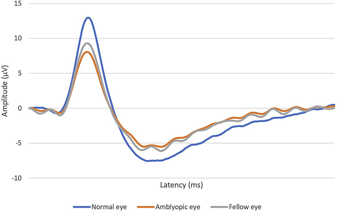
The refractive error found in the NE group of subjects had a mean of - 0.95 ± 1.65D, while the amblyopic group showed a mean of - 2.03 ± 4.29D. The VA in amblyopic eyes had a mean of 0.38 ± 0.20 logMAR. Analyzing PERG data, we observed significant differences in the P50-N95 amplitudes of the amblyopic group compared with the NE group (p < 0.0001-amblyopic eye vs. NE; p = 0.039-fellow eye vs. NE).

DISCUSSION

These findings suggest that amblyopic patients may also present other impairments beyond the visual cortex. PERGs seem to be an important complementary examination in the diagnosis of other impairments in amblyopia.

摘要

引言

弱视通常是单侧障碍,定义为双眼最佳矫正视力相差至少两行,对比度敏感度下降,以及立体视下降。图形视网膜电图(PERG)是一种非侵入性技术,提供视网膜生物电位,是黄斑区变化的高度敏感指标。我们的目的是评估弱视眼与正常眼(NE)的视网膜反应是否存在差异。

方法

我们评估了 24 名成年志愿者,12 名弱视患者(平均年龄 43.42±12.72 岁)和 12 名 NE 患者(平均年龄 35.58±12.85 岁)。两组受试者均无合并症。进行了全面的眼科检查,包括远距和近距视力(ETDRS 图表)、眼球对齐(遮盖试验)和 PERG 评估视网膜细胞反应等参数。

结果

NE 组受试者的屈光不正平均值为-0.95±1.65D,而弱视组则显示平均值为-2.03±4.29D。弱视眼的视力平均值为 0.38±0.20 logMAR。分析 PERG 数据,我们观察到弱视组的 P50-N95 振幅与 NE 组有显著差异(p<0.0001-弱视眼与 NE 组;p=0.039-对侧眼与 NE 组)。

讨论

这些发现表明,弱视患者可能还存在其他视觉皮层以外的损伤。PERG 似乎是弱视诊断中其他损伤的重要补充检查。

https://cdn.ncbi.nlm.nih.gov/pmc/blobs/694b/10873232/369f0f308afc/10792_2024_3042_Fig2_HTML.jpg
https://cdn.ncbi.nlm.nih.gov/pmc/blobs/694b/10873232/75414f126656/10792_2024_3042_Fig1_HTML.jpg
https://cdn.ncbi.nlm.nih.gov/pmc/blobs/694b/10873232/369f0f308afc/10792_2024_3042_Fig2_HTML.jpg
https://cdn.ncbi.nlm.nih.gov/pmc/blobs/694b/10873232/75414f126656/10792_2024_3042_Fig1_HTML.jpg
https://cdn.ncbi.nlm.nih.gov/pmc/blobs/694b/10873232/369f0f308afc/10792_2024_3042_Fig2_HTML.jpg

相似文献

1
Pattern electroretinography response in amblyopic adults.弱视成人的图形视网膜电图反应。
Int Ophthalmol. 2024 Feb 16;44(1):86. doi: 10.1007/s10792-024-03042-8.
2
Can pattern electroretinography be a relevant diagnostic aid in amblyopia? - A systematic review.视诱发电位图在弱视诊断中的作用:系统评价
Semin Ophthalmol. 2022 Jul 4;37(5):593-601. doi: 10.1080/08820538.2022.2069471. Epub 2022 May 6.
3
Long-term changes in visual acuity and refractive error in amblyopes.弱视患者视力和屈光不正的长期变化
Optom Vis Sci. 2004 Jul;81(7):510-5. doi: 10.1097/00006324-200407000-00012.
4
Multifocal electroretinography in amblyopia.多焦视网膜电图在弱视中的应用。
Graefes Arch Clin Exp Ophthalmol. 2020 Mar;258(3):683-691. doi: 10.1007/s00417-019-04558-x. Epub 2020 Jan 3.
5
Pattern visual evoked potential and foveal sensitivity in amblyopia.弱视的图形视觉诱发电位和中心凹敏感性。
Doc Ophthalmol. 2023 Oct;147(2):109-119. doi: 10.1007/s10633-023-09948-5. Epub 2023 Aug 28.
6
Binocular versus standard occlusion or blurring treatment for unilateral amblyopia in children aged three to eight years.双眼视刺激或常规遮盖治疗法治疗 3-8 岁儿童单眼弱视的比较。
Cochrane Database Syst Rev. 2022 Feb 7;2(2):CD011347. doi: 10.1002/14651858.CD011347.pub3.
7
Analysis of retinal nerve fiber layer thickness in anisometropic amblyopia via optic coherence tomography.通过光学相干断层扫描分析屈光参差性弱视患者的视网膜神经纤维层厚度
Graefes Arch Clin Exp Ophthalmol. 2019 Oct;257(10):2103-2110. doi: 10.1007/s00417-019-04402-2. Epub 2019 Jul 4.
8
Electrophysiological detection of delayed postretinal neural conduction in human amblyopia.人类弱视视网膜后神经传导延迟的电生理检测
Invest Ophthalmol Vis Sci. 2010 Oct;51(10):5041-8. doi: 10.1167/iovs.10-5412. Epub 2010 May 12.
9
A morphological study of amblyopic eyes in children failing to achieve normal visual acuity after electronically monitored long-term occlusion treatment.对经电子监测长期遮盖治疗后视力未达到正常的儿童弱视眼进行的形态学研究。
Graefes Arch Clin Exp Ophthalmol. 2015 Nov;253(11):2021-8. doi: 10.1007/s00417-015-3085-0. Epub 2015 Jun 23.
10
Characterising the orientation-specific pattern-onset visual evoked potentials in children with bilateral refractive amblyopia and non-amblyopic controls.描述双侧屈光性弱视儿童与非弱视对照者的视诱发电位朝向特异性图形翻转。
Doc Ophthalmol. 2021 Apr;142(2):197-211. doi: 10.1007/s10633-020-09794-9. Epub 2020 Sep 23.

本文引用的文献

1
Axial Length in Patients with Myopia and Interpretation of Pattern Electroretinogram Recordings.近视患者的眼轴长度及图形视网膜电图记录解读
Clin Ophthalmol. 2021 Jun 29;15:2815-2821. doi: 10.2147/OPTH.S308604. eCollection 2021.
2
Transcranial Magnetic Stimulation in Adults With Amblyopia.经颅磁刺激治疗成人弱视。
J Neuroophthalmol. 2020 Jun;40(2):185-192. doi: 10.1097/WNO.0000000000000828.
3
Prevalence and Risk Factors of Amblyopia among Refractive Errors in an Eastern European Population.东欧人群屈光不正性弱视的患病率及危险因素。
Medicina (Kaunas). 2018 Mar 20;54(1):6. doi: 10.3390/medicina54010006.
4
Retinothalamic White Matter Abnormalities in Amblyopia.弱视的视放射白质异常。
Invest Ophthalmol Vis Sci. 2018 Feb 1;59(2):921-929. doi: 10.1167/iovs.17-22930.
5
The Puzzle of Visual Development: Behavior and Neural Limits.视觉发育之谜:行为与神经限制
J Neurosci. 2016 Nov 9;36(45):11384-11393. doi: 10.1523/JNEUROSCI.2937-16.2016.
6
Steady-state pattern electroretinogram and frequency doubling technology in anisometropic amblyopia.屈光参差性弱视的稳态图形视网膜电图和频率加倍技术
Clin Ophthalmol. 2016 Oct 19;10:2061-2068. doi: 10.2147/OPTH.S117803. eCollection 2016.
7
Short-term effects of vision trainer rehabilitation in patients affected by anisometropic amblyopia: electrofunctional evaluation.屈光参差性弱视患者视觉训练康复的短期效果:电功能评估
Doc Ophthalmol. 2014 Dec;129(3):177-89. doi: 10.1007/s10633-014-9462-x. Epub 2014 Oct 8.
8
Advance in ERG analysis: from peak time and amplitude to frequency, power, and energy.ERG 分析的进展:从峰时和振幅到频率、功率和能量。
Biomed Res Int. 2014;2014:246096. doi: 10.1155/2014/246096. Epub 2014 Jul 1.
9
A morphological study of retinal changes in unilateral amblyopia using optical coherence tomography image segmentation.利用光学相干断层扫描图像分割技术对单侧弱视视网膜变化进行的形态学研究。
PLoS One. 2014 Feb 6;9(2):e88363. doi: 10.1371/journal.pone.0088363. eCollection 2014.
10
Structural and functional comparison of the persistent and resolved amblyopia.持续性弱视与恢复性弱视的结构和功能比较。
Doc Ophthalmol. 2014 Apr;128(2):101-9. doi: 10.1007/s10633-013-9422-x. Epub 2013 Dec 18.