Los Jonathan, Mensink Frans B, Mohammadnia Niekbachsh, Opstal Tjerk S J, Damman Peter, Volleberg Rick H J A, Peeters Denise A M, van Royen Niels, Garcia-Garcia Hector M, Cornel Jan H, El Messaoudi Saloua, van Geuns Robert-Jan M
Department of Cardiology, Radboud University Medical Center, Nijmegen, Netherlands.
Department of Cardiology, Northwest Clinics, Alkmaar, Netherlands.
Front Cardiovasc Med. 2024 Feb 1;11:1352025. doi: 10.3389/fcvm.2024.1352025. eCollection 2024.
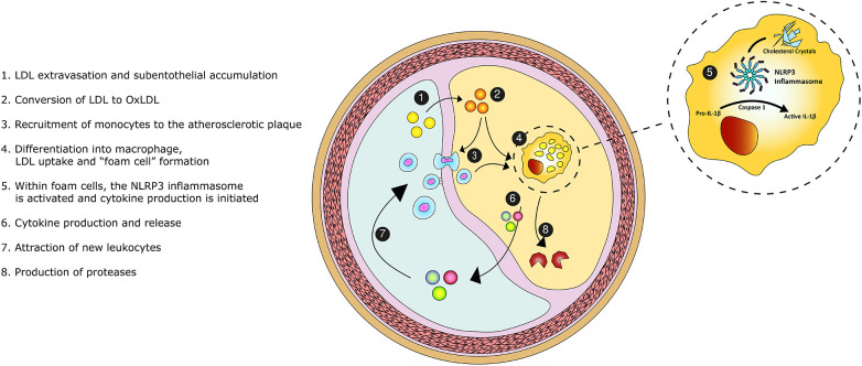
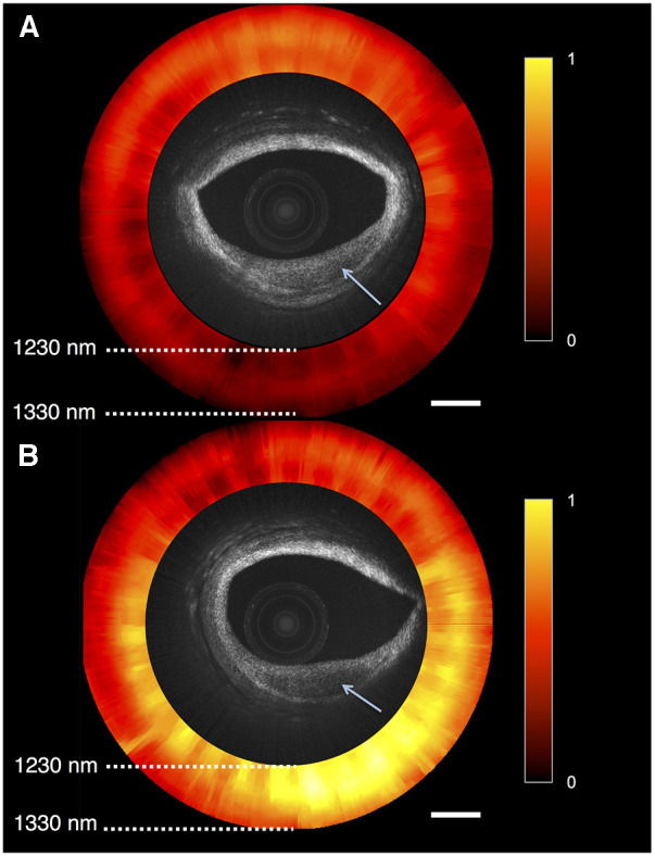
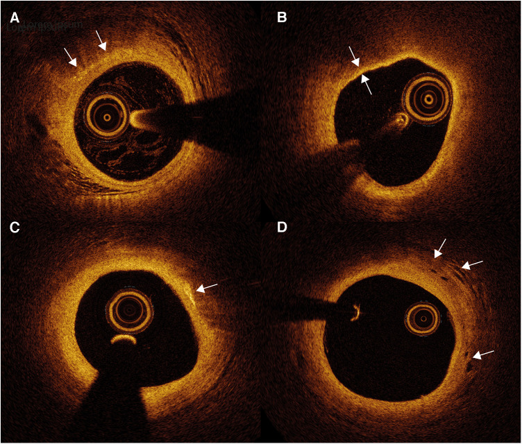
Coronary atherosclerosis remains a leading cause of morbidity and mortality worldwide. The underlying pathophysiology includes a complex interplay of endothelial dysfunction, lipid accumulation and inflammatory pathways. Multiple structural and inflammatory features of the atherosclerotic lesions have become targets to identify high-risk lesions. Various intracoronary imaging devices have been developed to assess the morphological, biocompositional and molecular profile of the intracoronary atheromata. These techniques guide interventional and therapeutical management and allow the identification and stratification of atherosclerotic lesions. We sought to provide an overview of the inflammatory pathobiology of atherosclerosis, distinct high-risk plaque features and the ability to visualize this process with contemporary intracoronary imaging techniques.
冠状动脉粥样硬化仍然是全球发病和死亡的主要原因。其潜在的病理生理学包括内皮功能障碍、脂质积聚和炎症途径之间复杂的相互作用。动脉粥样硬化病变的多种结构和炎症特征已成为识别高危病变的靶点。已开发出各种冠状动脉内成像设备,以评估冠状动脉粥样斑块的形态、生物成分和分子特征。这些技术指导介入和治疗管理,并有助于识别动脉粥样硬化病变并对其进行分层。我们试图概述动脉粥样硬化的炎症病理生物学、独特的高危斑块特征以及使用当代冠状动脉内成像技术可视化这一过程的能力。