Department of Anatomy & Embryology, Faculty of Health, Medicine and Life Sciences, Maastricht University, Universiteitssingel 50, 6229 ER, Maastricht, The Netherlands.
Tytgat Institute for Liver and Intestinal Research, Academic Medical Center, University of Amsterdam, Amsterdam, The Netherlands.
Clin Auton Res. 2024 Feb;34(1):79-97. doi: 10.1007/s10286-024-01023-6. Epub 2024 Feb 25.
We have re-evaluated the anatomical arguments that underlie the division of the spinal visceral outflow into sympathetic and parasympathetic divisions.
Using a systematic literature search, we mapped the location of catecholaminergic neurons throughout the mammalian peripheral nervous system. Subsequently, a narrative method was employed to characterize segment-dependent differences in the location of preganglionic cell bodies and the composition of white and gray rami communicantes.
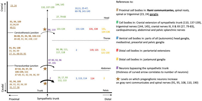
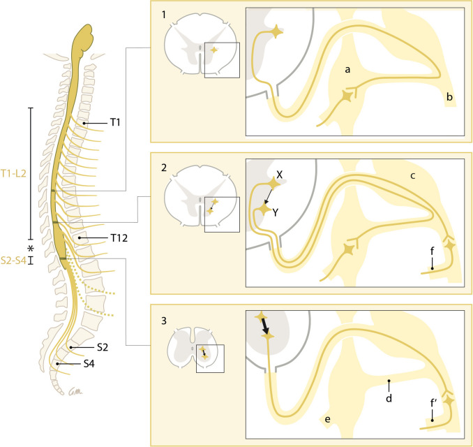
One hundred seventy studies were included in the systematic review, providing information on 389 anatomical structures. Catecholaminergic nerve fibers are present in most spinal and all cranial nerves and ganglia, including those that are known for their parasympathetic function. Along the entire spinal autonomic outflow pathways, proximal and distal catecholaminergic cell bodies are common in the head, thoracic, and abdominal and pelvic region, which invalidates the "short-versus-long preganglionic neuron" argument. Contrary to the classically confined outflow levels T1-L2 and S2-S4, preganglionic neurons have been found in the resulting lumbar gap. Preganglionic cell bodies that are located in the intermediolateral zone of the thoracolumbar spinal cord gradually nest more ventrally within the ventral motor nuclei at the lumbar and sacral levels, and their fibers bypass the white ramus communicans and sympathetic trunk to emerge directly from the spinal roots. Bypassing the sympathetic trunk, therefore, is not exclusive for the sacral outflow. We conclude that the autonomic outflow displays a conserved architecture along the entire spinal axis, and that the perceived differences in the anatomy of the autonomic thoracolumbar and sacral outflow are quantitative.
我们重新评估了支配脊髓内脏传出分为交感和副交感两部分的解剖学论点。
通过系统文献检索,我们绘制了哺乳动物周围神经系统中儿茶酚胺能神经元的位置图。随后,采用叙述性方法来描述节前细胞体在节段上的位置以及白交通支和灰交通支的组成的差异。
系统综述共纳入 170 项研究,提供了 389 个解剖结构的信息。儿茶酚胺能神经纤维存在于大多数脊神经和所有颅神经和神经节中,包括那些以副交感功能而闻名的神经节。沿着整个脊髓自主传出通路,头、胸和腹盆区近端和远端的儿茶酚胺能细胞体都很常见,这否定了“短节前神经元与长节前神经元”的论点。与经典的 T1-L2 和 S2-S4 传出水平相反,在由此产生的腰区间隙中发现了节前神经元。位于胸腰椎脊髓中间外侧区的节前细胞体逐渐向腹侧在腰骶水平的腹运动核内嵌套,其纤维绕过白交通支和交感干,直接从脊神经根发出。因此,绕过交感干并不是骶部传出的特有现象。我们的结论是,自主传出在整个脊髓轴上具有保守的结构,而人们对自主胸腰椎和骶部传出的解剖差异的认识是定量的。