Department of Anatomy & Embryology, Maastricht University, Maastricht, The Netherlands.
Department of Anatomy, Faculty of Science, Mahidol University, Bangkok, Thailand.
J Anat. 2020 Oct;237(4):655-671. doi: 10.1111/joa.13230. Epub 2020 Jun 29.
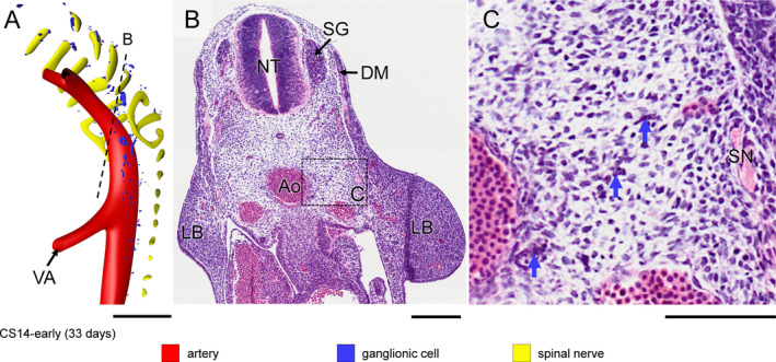
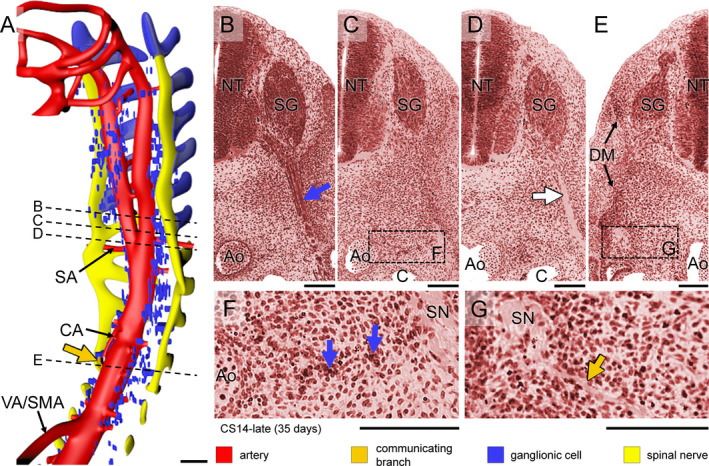
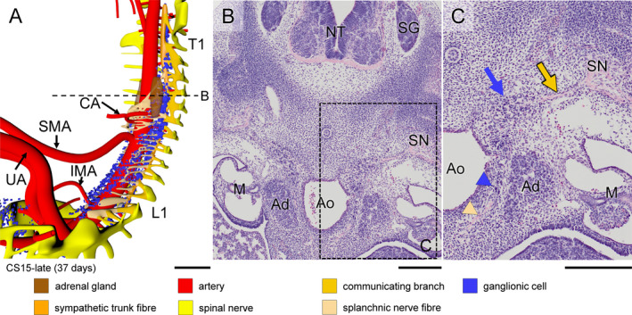
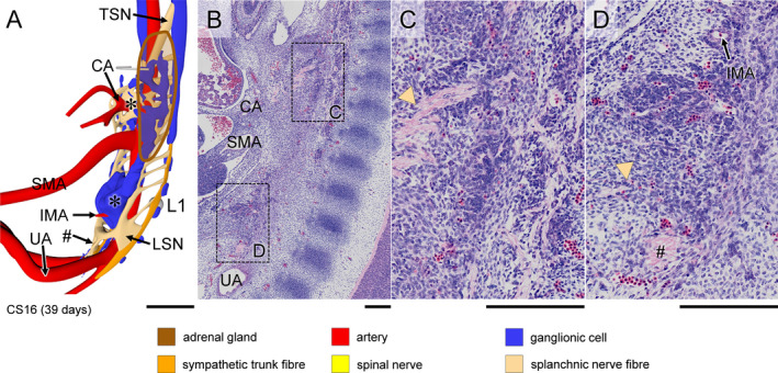
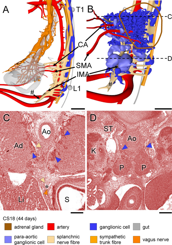
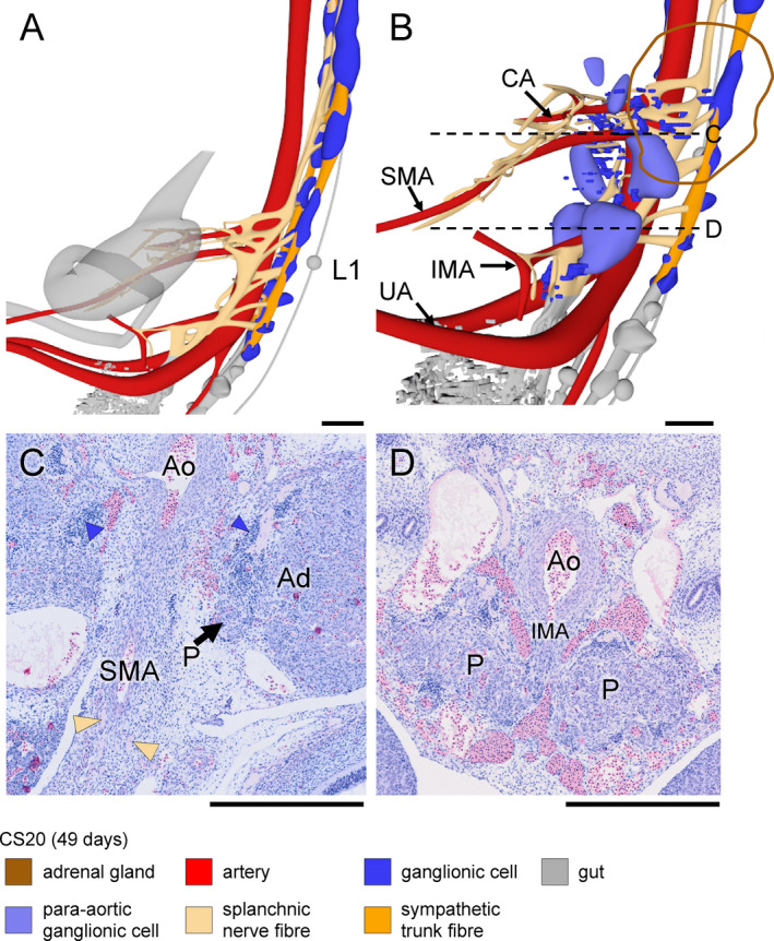
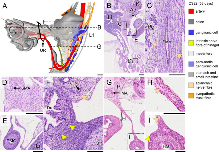
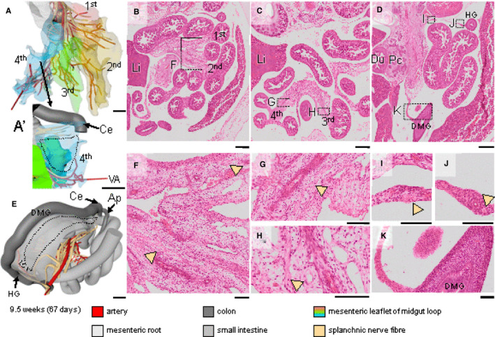
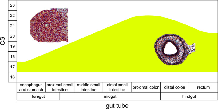
Compared to the intrinsic enteric nervous system (ENS), development of the extrinsic ENS is poorly documented, even though its presence is easily detectable with histological techniques. We visualised its development in human embryos and foetuses of 4-9.5 weeks post-fertilisation using Amira 3D-reconstruction and Cinema 4D-remodelling software. The extrinsic ENS originated from small, basophilic neural crest cells (NCCs) that migrated to the para-aortic region and then continued ventrally to the pre-aortic region, where they formed autonomic pre-aortic plexuses. From here, nerve fibres extended along the ventral abdominal arteries and finally connected to the intrinsic system. Schwann cell precursors (SCPs), a subgroup of NCCs that migrate on nerve fibres, showed region-specific differences in differentiation. SCPs developed into scattered chromaffin cells of the adrenal medulla dorsolateral to the coeliac artery (CA) and into more tightly packed chromaffin cells of the para-aortic bodies ventrolateral to the inferior mesenteric artery (IMA), with reciprocal topographic gradients between both fates. The extrinsic ENS first extended along the CA and then along the superior mesenteric artery (SMA) and IMA 5 days later. Apart from the branch to the caecum, extrinsic nerves did not extend along SMA branches in the herniated parts of the midgut until the gut loops had returned in the abdominal cavity, suggesting a permissive role of the intraperitoneal environment. Accordingly, extrinsic innervation had not yet reached the distal (colonic) loop of the midgut at 9.5 weeks development. Based on intrinsic ENS-dependent architectural remodelling of the gut layers, extrinsic innervation followed intrinsic innervation 3-4 Carnegie stages later.
与内在肠神经系统(ENS)相比,外在 ENS 的发育过程记录较差,尽管其存在很容易通过组织学技术检测到。我们使用 Amira 3D 重建和 Cinema 4D 重塑软件在人类胚胎和受精后 4-9.5 周的胎儿中观察到其发育情况。外在 ENS 起源于小的嗜碱性神经嵴细胞(NCC),它们迁移到腹主动脉旁区域,然后继续向腹侧迁移到主动脉前区域,在那里形成自主主动脉前丛。从这里,神经纤维沿着腹主动脉延伸,最终与内在系统连接。Schwann 细胞前体(SCP)是迁移到神经纤维上的 NCC 的一个亚群,在分化方面表现出区域特异性差异。SCP 发育成腹主动脉旁体的散在嗜铬细胞,位于腹腔动脉(CA)的背外侧,以及腹主动脉下体的更紧密排列的嗜铬细胞,两者之间存在相互的地形梯度。外在 ENS 首先沿着 CA 延伸,然后在 5 天后沿着肠系膜上动脉(SMA)和肠系膜下动脉(IMA)延伸。除了通向盲肠的分支外,外在神经在中肠疝出部分的 SMA 分支上没有延伸,直到肠环返回腹腔,这表明腹腔内环境具有允许作用。因此,外在神经支配在 9.5 周发育时还没有到达中肠的远端(结肠)环。基于内在 ENS 依赖的肠层结构重塑,外在神经支配在内在神经支配后 3-4 个 Carnegie 阶段后出现。