Merckel L G, Pomp J, Hackett S L, van Lier A L H M W, van den Dobbelsteen M, Rasing M J A, Mohamed Hoesein F A A, Snoeren L M W, van Es C A, van Rossum P S N, Fast M F, Verhoeff J J C
Department of Radiotherapy, University Medical Center Utrecht, Heidelberglaan 100, 3584 CX Utrecht, The Netherlands.
Department of Radiology, University Medical Center Utrecht, Utrecht, The Netherlands.
Clin Transl Radiat Oncol. 2024 Feb 15;45:100744. doi: 10.1016/j.ctro.2024.100744. eCollection 2024 Mar.
MRI-guidance may aid better discrimination between Organs at Risk (OARs) and target volumes in proximity of the mediastinum. We report the first clinical experiences with Stereotactic Body Radiotherapy (SBRT) of (ultra)central lung tumours on a 1.5 T MR-linac.
Patients with an (ultra)central lung tumour were selected for MR-linac based SBRT treatment. A T2-weighted 3D sequence MRI acquired during free breathing was used for daily plan adaption. Prior to each fraction, contours of Internal Target Volume (ITV) and OARs were deformably propagated and amended by a radiation oncologist. Inter-fractional changes in volumes and coverage of target volumes as well as doses in OARs were evaluated in offline and online treatment plans.
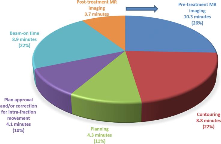
Ten patients were treated and completed 60 Gy in 8 or 12 fractions. In total 104 fractions were delivered. The median time in the treatment room was 41 min with a median beam-on time of 8.9 min. No grade ≥3 acute toxicity was observed. In two patients, the ITV significantly decreased during treatment (58 % and 37 %, respectively) due to tumour shrinkage. In the other patients, 81 % of online ITVs were within ±15 % of the volume of fraction 1. Comparison with the pre-treatment plan showed that ITV coverage of the online plan was similar in 52 % and improved in 34 % of cases. Adaptation to meet OAR constraints, led to decreased ITV coverage in 14 %.
We describe the workflow for MR-guided Radiotherapy and the feasibility of using 1.5 T MR-linac for SBRT of (ultra) central lung tumours.
磁共振成像(MRI)引导有助于更好地区分危险器官(OARs)与纵隔附近的靶区。我们报告了在1.5T MR直线加速器上对(超)中央型肺肿瘤进行立体定向体部放疗(SBRT)的首例临床经验。
选择(超)中央型肺肿瘤患者接受基于MR直线加速器的SBRT治疗。在自由呼吸期间采集的T2加权3D序列MRI用于每日计划调整。在每个分次治疗前,由放射肿瘤学家对内部靶区(ITV)和OARs的轮廓进行可变形传播和修正。在离线和在线治疗计划中评估靶区体积和覆盖范围以及OARs剂量的分次间变化。
10例患者接受治疗,分8次或12次完成60Gy照射。共进行了104次分次治疗。在治疗室的中位时间为41分钟,中位照射时间为8.9分钟。未观察到≥3级急性毒性反应。在2例患者中,由于肿瘤缩小,治疗期间ITV显著减小(分别为58%和37%)。在其他患者中,81%的在线ITV在第1分次体积的±15%范围内。与治疗前计划相比,在线计划的ITV覆盖在52%的病例中相似,在34%的病例中得到改善。为满足OAR限制而进行的调整导致14%的病例中ITV覆盖降低。
我们描述了MR引导放疗的工作流程以及使用1.5T MR直线加速器对(超)中央型肺肿瘤进行SBRT的可行性。