• 文献检索
  • 文档翻译
  • 深度研究
  • 学术资讯
  • Suppr Zotero 插件Zotero 插件
  • 邀请有礼
  • 套餐&价格
  • 历史记录
应用&插件
Suppr Zotero 插件Zotero 插件浏览器插件Mac 客户端Windows 客户端微信小程序
定价
高级版会员购买积分包购买API积分包
服务
文献检索文档翻译深度研究API 文档MCP 服务
关于我们
关于 Suppr公司介绍联系我们用户协议隐私条款
关注我们

Suppr 超能文献

核心技术专利:CN118964589B侵权必究
粤ICP备2023148730 号-1Suppr @ 2026

文献检索

告别复杂PubMed语法,用中文像聊天一样搜索,搜遍4000万医学文献。AI智能推荐,让科研检索更轻松。

立即免费搜索

文件翻译

保留排版,准确专业,支持PDF/Word/PPT等文件格式,支持 12+语言互译。

免费翻译文档

深度研究

AI帮你快速写综述,25分钟生成高质量综述,智能提取关键信息,辅助科研写作。

立即免费体验

溶酶体(功能异常)在动脉粥样硬化中的作用——小细胞器肩负重任

Lysosome (Dys)function in Atherosclerosis-A Big Weight on the Shoulders of a Small Organelle.

作者信息

Marques André R A, Ramos Cristiano, Machado-Oliveira Gisela, Vieira Otília V

机构信息

iNOVA4Health, Chronic Diseases Research Center (CEDOC), NOVA Medical School (NMS), Universidade NOVA de Lisboa, Lisbon, Portugal.

出版信息

Front Cell Dev Biol. 2021 Mar 29;9:658995. doi: 10.3389/fcell.2021.658995. eCollection 2021.

DOI:10.3389/fcell.2021.658995
PMID:33855029
原文链接:https://pmc.ncbi.nlm.nih.gov/articles/PMC8039146/
Abstract

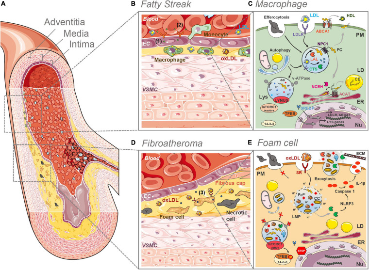
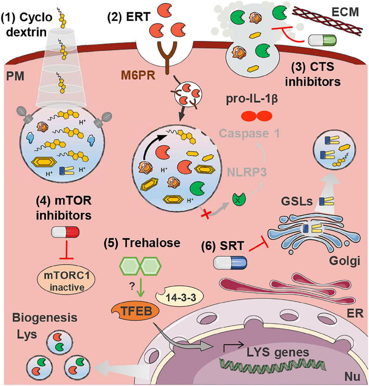
Atherosclerosis is a progressive insidious chronic disease that underlies most of the cardiovascular pathologies, including myocardial infarction and ischemic stroke. The malfunctioning of the lysosomal compartment has a central role in the etiology and pathogenesis of atherosclerosis. Lysosomes are the degradative organelles of mammalian cells and process endogenous and exogenous substrates in a very efficient manner. Dysfunction of these organelles and consequent inefficient degradation of modified low-density lipoproteins (LDL) and apoptotic cells in atherosclerotic lesions have, therefore, numerous deleterious consequences for cellular homeostasis and disease progression. Lysosome dysfunction has been mostly studied in the context of the inherited lysosomal storage disorders (LSDs). However, over the last years it has become increasingly evident that the consequences of this phenomenon are more far-reaching, also influencing the progression of multiple acquired human pathologies, such as neurodegenerative diseases, cancer, and cardiovascular diseases (CVDs). During the formation of atherosclerotic plaques, the lysosomal compartment of the various cells constituting the arterial wall is under severe stress, due to the tremendous amounts of lipoproteins being processed by these cells. The uncontrolled uptake of modified lipoproteins by arterial phagocytic cells, namely macrophages and vascular smooth muscle cells (VSMCs), is the initial step that triggers the pathogenic cascade culminating in the formation of atheroma. These cells become pathogenic "foam cells," which are characterized by dysfunctional lipid-laden lysosomes. Here, we summarize the current knowledge regarding the origin and impact of the malfunctioning of the lysosomal compartment in plaque cells. We further analyze how the field of LSD research may contribute with some insights to the study of CVDs, particularly how therapeutic approaches that target the lysosomes in LSDs could be applied to hamper atherosclerosis progression and associated mortality.

摘要

动脉粥样硬化是一种渐进性、隐匿性的慢性疾病,是大多数心血管疾病的基础,包括心肌梗死和缺血性中风。溶酶体区室功能异常在动脉粥样硬化的病因和发病机制中起核心作用。溶酶体是哺乳动物细胞的降解细胞器,能非常高效地处理内源性和外源性底物。因此,这些细胞器功能障碍以及动脉粥样硬化病变中修饰的低密度脂蛋白(LDL)和凋亡细胞降解效率低下,对细胞稳态和疾病进展产生了许多有害影响。溶酶体功能障碍大多是在遗传性溶酶体贮积症(LSD)的背景下进行研究的。然而,在过去几年中越来越明显的是,这种现象的后果更为深远,也影响多种后天性人类疾病的进展,如神经退行性疾病、癌症和心血管疾病(CVD)。在动脉粥样硬化斑块形成过程中,构成动脉壁的各种细胞的溶酶体区室承受着巨大压力,因为这些细胞要处理大量脂蛋白。动脉吞噬细胞,即巨噬细胞和血管平滑肌细胞(VSMC)对修饰脂蛋白的不受控制摄取是引发致病级联反应的第一步,最终导致动脉粥样瘤形成。这些细胞变成致病性“泡沫细胞”,其特征是充满脂质的溶酶体功能失调。在此,我们总结了关于斑块细胞中溶酶体区室功能异常的起源和影响的现有知识。我们进一步分析LSD研究领域如何能为CVD研究提供一些见解,特别是针对LSD中溶酶体的治疗方法如何应用于阻碍动脉粥样硬化进展和相关死亡率。

https://cdn.ncbi.nlm.nih.gov/pmc/blobs/0f63/8039146/427b324e099f/fcell-09-658995-g002.jpg
https://cdn.ncbi.nlm.nih.gov/pmc/blobs/0f63/8039146/765d6ed99e2e/fcell-09-658995-g001.jpg
https://cdn.ncbi.nlm.nih.gov/pmc/blobs/0f63/8039146/427b324e099f/fcell-09-658995-g002.jpg
https://cdn.ncbi.nlm.nih.gov/pmc/blobs/0f63/8039146/765d6ed99e2e/fcell-09-658995-g001.jpg
https://cdn.ncbi.nlm.nih.gov/pmc/blobs/0f63/8039146/427b324e099f/fcell-09-658995-g002.jpg

相似文献

1
Lysosome (Dys)function in Atherosclerosis-A Big Weight on the Shoulders of a Small Organelle.溶酶体(功能异常)在动脉粥样硬化中的作用——小细胞器肩负重任
Front Cell Dev Biol. 2021 Mar 29;9:658995. doi: 10.3389/fcell.2021.658995. eCollection 2021.
2
Cholesteryl hemiazelate causes lysosome dysfunction impacting vascular smooth muscle cell homeostasis.胆甾醇半缩醛酯导致溶酶体功能障碍,影响血管平滑肌细胞的动态平衡。
J Cell Sci. 2022 Mar 1;135(5). doi: 10.1242/jcs.254631. Epub 2021 Oct 22.
3
Cholesteryl Hemiazelate Present in Cardiovascular Disease Patients Causes Lysosome Dysfunction in Murine Fibroblasts.胆固醇酰基溶血磷脂酸存在于心血管疾病患者中,导致小鼠成纤维细胞溶酶体功能障碍。
Cells. 2023 Dec 13;12(24):2826. doi: 10.3390/cells12242826.
4
Autophagy and Lysosome Storage Disorders.自噬与溶酶体贮积症
Adv Exp Med Biol. 2020;1207:87-102. doi: 10.1007/978-981-15-4272-5_5.
5
Degradation and beyond: the macrophage lysosome as a nexus for nutrient sensing and processing in atherosclerosis.降解及其他:巨噬细胞溶酶体作为动脉粥样硬化中营养感知与处理的枢纽
Curr Opin Lipidol. 2015 Oct;26(5):394-404. doi: 10.1097/MOL.0000000000000213.
6
Lysosomal acid lipase: at the crossroads of normal and atherogenic cholesterol metabolism.溶酶体酸性脂肪酶:在正常和动脉粥样硬化胆固醇代谢的十字路口。
Front Cell Dev Biol. 2015 Feb 2;3:3. doi: 10.3389/fcell.2015.00003. eCollection 2015.
7
Role of lysosomes in physiological activities, diseases, and therapy.溶酶体在生理活动、疾病和治疗中的作用。
J Hematol Oncol. 2021 May 14;14(1):79. doi: 10.1186/s13045-021-01087-1.
8
[Does Lp-PLA2 determination help predict atherosclerosis and cardiocerebrovascular disease?].脂蛋白磷脂酶A2测定有助于预测动脉粥样硬化和心脑血管疾病吗?
Acta Med Croatica. 2010 Oct;64(4):237-45.
9
eIF2α-mediated integrated stress response links multiple intracellular signaling pathways to reprogram vascular smooth muscle cell fate in carotid artery plaque.真核生物起始因子2α(eIF2α)介导的综合应激反应将多条细胞内信号通路与颈动脉斑块中血管平滑肌细胞命运重编程联系起来。
Heliyon. 2024 Feb 23;10(5):e26904. doi: 10.1016/j.heliyon.2024.e26904. eCollection 2024 Mar 15.
10
The role of microscopy in understanding atherosclerotic lysosomal lipid metabolism.显微镜检查在理解动脉粥样硬化溶酶体脂质代谢中的作用。
Microsc Microanal. 2003 Feb;9(1):54-67. doi: 10.1017/S1431927603030010.

引用本文的文献

1
Glucosylated cholesterol accumulates in atherosclerotic lesions and impacts macrophage immune response.糖基化胆固醇在动脉粥样硬化病变中蓄积,并影响巨噬细胞免疫反应。
J Lipid Res. 2025 Jun;66(6):100825. doi: 10.1016/j.jlr.2025.100825. Epub 2025 May 15.
2
Down-regulation of ATP8B2 in Foam Cells Inhibits Autophagic Flux and ox-LDL Degradation in Atherosclerosis.泡沫细胞中ATP8B2的下调抑制动脉粥样硬化中的自噬通量和氧化型低密度脂蛋白降解。
Cell Biochem Biophys. 2025 Mar 28. doi: 10.1007/s12013-025-01728-z.
3
Lysosome Functions in Atherosclerosis: A Potential Therapeutic Target.

本文引用的文献

1
Macrophage SR-BI modulates autophagy via VPS34 complex and PPARα transcription of Tfeb in atherosclerosis.巨噬细胞SR-BI通过VPS34复合物和PPARα转录Tfeb调节动脉粥样硬化中的自噬。
J Clin Invest. 2021 Apr 1;131(7). doi: 10.1172/JCI94229.
2
Lysosomal SLC46A3 modulates hepatic cytosolic copper homeostasis.溶酶体 SLC46A3 调节肝脏细胞质内铜稳态。
Nat Commun. 2021 Jan 12;12(1):290. doi: 10.1038/s41467-020-20461-0.
3
From Inert Storage to Biological Activity-In Search of Identity for Oxidized Cholesteryl Esters.从惰性储存到生物活性——探寻氧化胆固醇酯的真面目
溶酶体在动脉粥样硬化中的功能:一个潜在的治疗靶点。
Cells. 2025 Jan 24;14(3):183. doi: 10.3390/cells14030183.
4
Haematometabolism rewiring in atherosclerotic cardiovascular disease.动脉粥样硬化性心血管疾病中的血液代谢重塑
Nat Rev Cardiol. 2025 Jun;22(6):414-430. doi: 10.1038/s41569-024-01108-9. Epub 2025 Jan 2.
5
Cholesterol crystals in the pathogenesis of atherosclerosis.胆固醇晶体在动脉粥样硬化发病机制中的作用
Nat Rev Cardiol. 2025 May;22(5):315-332. doi: 10.1038/s41569-024-01100-3. Epub 2024 Nov 18.
6
Phenotypic, Metabolic, and Functional Characterization of Experimental Models of Foamy Macrophages: Toward Therapeutic Research in Atherosclerosis.泡沫巨噬细胞实验模型的表型、代谢和功能特征:动脉粥样硬化治疗研究的进展。
Int J Mol Sci. 2024 Sep 21;25(18):10146. doi: 10.3390/ijms251810146.
7
Lysosomes in the immunometabolic reprogramming of immune cells in atherosclerosis.溶酶体在动脉粥样硬化中免疫细胞的免疫代谢重编程过程中的作用
Nat Rev Cardiol. 2025 Mar;22(3):149-164. doi: 10.1038/s41569-024-01072-4. Epub 2024 Sep 20.
8
eIF2α-mediated integrated stress response links multiple intracellular signaling pathways to reprogram vascular smooth muscle cell fate in carotid artery plaque.真核生物起始因子2α(eIF2α)介导的综合应激反应将多条细胞内信号通路与颈动脉斑块中血管平滑肌细胞命运重编程联系起来。
Heliyon. 2024 Feb 23;10(5):e26904. doi: 10.1016/j.heliyon.2024.e26904. eCollection 2024 Mar 15.
9
Cholesteryl Hemiazelate Present in Cardiovascular Disease Patients Causes Lysosome Dysfunction in Murine Fibroblasts.胆固醇酰基溶血磷脂酸存在于心血管疾病患者中,导致小鼠成纤维细胞溶酶体功能障碍。
Cells. 2023 Dec 13;12(24):2826. doi: 10.3390/cells12242826.
10
Bioinformatics analysis of immune cell infiltration patterns and potential diagnostic markers in atherosclerosis.动脉粥样硬化中免疫细胞浸润模式及潜在诊断标志物的生物信息学分析。
Sci Rep. 2023 Nov 14;13(1):19821. doi: 10.1038/s41598-023-47257-8.
Front Endocrinol (Lausanne). 2020 Nov 20;11:602252. doi: 10.3389/fendo.2020.602252. eCollection 2020.
4
NPC1-mTORC1 Signaling Couples Cholesterol Sensing to Organelle Homeostasis and Is a Targetable Pathway in Niemann-Pick Type C.NPC1-mTORC1 信号传导将胆固醇感应与细胞器动态平衡偶联起来,是尼曼-匹克 C 型的一个可靶向通路。
Dev Cell. 2021 Feb 8;56(3):260-276.e7. doi: 10.1016/j.devcel.2020.11.016. Epub 2020 Dec 11.
5
How Lysosomes Sense, Integrate, and Cope with Stress.溶酶体如何感知、整合和应对压力。
Trends Biochem Sci. 2021 Feb;46(2):97-112. doi: 10.1016/j.tibs.2020.09.004. Epub 2020 Oct 1.
6
Lysosomal quality control of cell fate: a novel therapeutic target for human diseases.溶酶体质量控制细胞命运:人类疾病的新治疗靶点。
Cell Death Dis. 2020 Sep 30;11(9):817. doi: 10.1038/s41419-020-03032-5.
7
Cell Senescence, Multiple Organelle Dysfunction and Atherosclerosis.细胞衰老、多细胞器功能障碍与动脉粥样硬化
Cells. 2020 Sep 23;9(10):2146. doi: 10.3390/cells9102146.
8
Impaired lipophagy in endothelial cells with prolonged exposure to oxidized low‑density lipoprotein.内皮细胞长期暴露于氧化型低密度脂蛋白可导致脂噬受损。
Mol Med Rep. 2020 Oct;22(4):2665-2672. doi: 10.3892/mmr.2020.11345. Epub 2020 Jul 16.
9
Metformin directly suppresses atherosclerosis in normoglycaemic mice via haematopoietic adenosine monophosphate-activated protein kinase.二甲双胍通过造血腺苷单磷酸激活蛋白激酶直接抑制血糖正常小鼠的动脉粥样硬化。
Cardiovasc Res. 2021 Apr 23;117(5):1295-1308. doi: 10.1093/cvr/cvaa171.
10
Cholesterol Handling in Lysosomes and Beyond.溶酶体及以外的胆固醇处理
Trends Cell Biol. 2020 Jun;30(6):452-466. doi: 10.1016/j.tcb.2020.02.007. Epub 2020 Mar 23.