Fan Wei, Sun Wei, Xu Ming Ze, Pan Jing Jing, Man Feng Yuan
Department of Radiology, Rocket Force Characteristic Medical Center of the Chinese People's Liberation Army, Beijing, China.
Department of Interventional Therapy, National Cancer Center/National Clinical Research Center for Cancer/Cancer Hospital, Chinese Academy of Medical Sciences and Peking Union Medical College, Beijing, China.
Front Oncol. 2024 Feb 21;14:1307907. doi: 10.3389/fonc.2024.1307907. eCollection 2024.
To establish a radiomics model for distinguishing between the benign and malignant mammary gland nodules via combining the features from nodule and mammary regions on DCE-MRI.
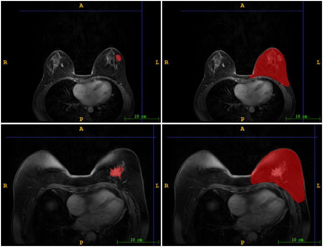
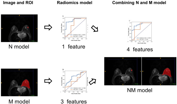
In this retrospective study, a total of 103 cases with mammary gland nodules (malignant/benign = 80/23) underwent DCE-MRI, and was confirmed by biopsy pathology. Features were extracted from both nodule region and mammary region on DCE-MRI. Three SVM classifiers were built for diagnosis of benign and malignant nodules as follows: the model with the features only from nodule region (N model), with the features only from mammary region (M model) and the model combining the features from nodule region and mammary region (NM model). The performance of models was evaluated with the area under the curve of receiver operating characteristic (AUC).
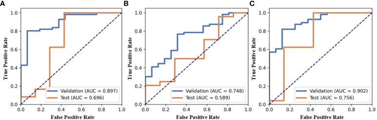
One radiomic features is selected from nodule region and 3 radiomic features is selected from mammary region. Compared with N or M model, NM model exhibited the best performance with an AUC of 0.756.
Compared with the model only using the features from nodule or mammary region, the radiomics-based model combining the features from nodule and mammary region outperformed in the diagnosis of benign and malignant nodules.
通过结合动态对比增强磁共振成像(DCE-MRI)上结节和乳腺区域的特征,建立一种区分乳腺良恶性结节的放射组学模型。
在这项回顾性研究中,共有103例乳腺结节患者(恶性/良性 = 80/23)接受了DCE-MRI检查,并经活检病理证实。从DCE-MRI上的结节区域和乳腺区域提取特征。构建了三个支持向量机(SVM)分类器用于诊断良性和恶性结节,具体如下:仅使用结节区域特征的模型(N模型)、仅使用乳腺区域特征的模型(M模型)以及结合结节区域和乳腺区域特征的模型(NM模型)。采用受试者操作特征曲线(ROC)下面积(AUC)评估模型性能。
从结节区域选择了1个放射组学特征,从乳腺区域选择了3个放射组学特征。与N模型或M模型相比,NM模型表现最佳,AUC为0.756。
与仅使用结节或乳腺区域特征的模型相比,结合结节和乳腺区域特征的基于放射组学的模型在诊断乳腺良恶性结节方面表现更优。