Bot Robert B, Chirla Razvan, Hozan Calin Tudor, Cavalu Simona
Faculty of Medicine and Pharmacy, University of Oradea, Oradea, 410087, Romania.
Department of Orthopedics, Emergency County Clinical Hospital Oradea, Oradea, 410169, Romania.
Int J Gen Med. 2024 Mar 21;17:1085-1100. doi: 10.2147/IJGM.S454546. eCollection 2024.
The purpose of this study was to quantify the modifications occurring in osteoporosis at the level of the human proximal femur throughout the trabecular structure, along with the identification of certain anatomic regions preferentially affected by osteoporosis. Another goal was to map the evolution of the radiodensity of the trabecular bone as osteoporosis progresses to an advanced stage.
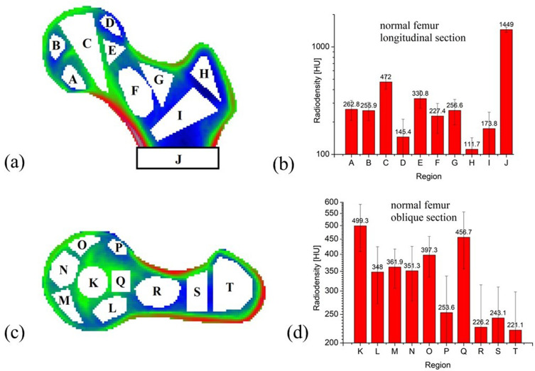
The study included CT scans (right femur) from 51 patients, out of which 40 had various degrees of osteoporosis, but no other local pathology. Ten regions of interest in two orthogonal slices have been identified and the differences in radiodensity as well as their evolution have been statistically analyzed in terms of relative and absolute changes.
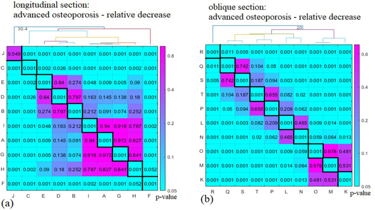
A detailed spatial map showing the evolution of osteoporosis was obtained. As osteoporosis evolved, the relative decrease in radiodensity was inversely correlated to the radiodensity of the healthy bone. In particular, the region covering the Ward triangle decreased the most, by an average 61-62% in osteopenia and 101-106% in advanced osteoporosis, while the principal compressive group was affected the least, showing a decrease by an average 14-15% in osteopenia and 29-32% in advanced osteoporosis. The absolute decrease in radiodensity was not correlated to the radiodensity of the healthy bone and was shifted to the inferior-posterior edge of the femur. Inside the femoral head, the upper region was affected the most in absolute terms, while the greater trochanter was less affected than the femoral neck. The maximum metaphyseal cortical bone density was unaffected by the progression of osteoporosis.
Significant differences were noticed in terms of the absolute and relative osteoporotic changes in radiodensity related to different anatomical regions of the human femoral bone. These differences become more pronounced as the disease progresses.
本研究的目的是量化人类近端股骨小梁结构层面骨质疏松症发生的变化,同时确定某些优先受骨质疏松症影响的解剖区域。另一个目标是绘制随着骨质疏松症进展到晚期小梁骨骨密度的变化情况。
该研究纳入了51例患者的CT扫描(右侧股骨),其中40例有不同程度的骨质疏松症,但无其他局部病变。在两个正交切片中确定了10个感兴趣区域,并对骨密度差异及其变化进行了相对和绝对变化方面的统计分析。
获得了一幅显示骨质疏松症变化的详细空间图。随着骨质疏松症的发展,骨密度的相对降低与健康骨的骨密度呈负相关。特别是,覆盖沃德三角区的区域减少最多,骨质减少时平均减少61%-62%,晚期骨质疏松症时减少101%-106%,而主要压缩组受影响最小,骨质减少时平均减少14%-15%,晚期骨质疏松症时减少29%-32%。骨密度的绝对降低与健康骨的骨密度无关,且转移至股骨的后下边缘。在股骨头内部,上部区域受影响的绝对值最大,而大转子比股骨颈受影响小。干骺端皮质骨最大密度不受骨质疏松症进展的影响。
在与人类股骨不同解剖区域相关的骨密度绝对和相对骨质疏松变化方面发现了显著差异。随着疾病进展,这些差异变得更加明显。