Wen Ru M, Qiu Zhengyuan, Marti G Edward W, Peterson Eric E, Marques Fernando Jose Garcia, Bermudez Abel, Wei Yi, Nolley Rosalie, Lam Nathan, Polasko Alex LaPat, Chiu Chun-Lung, Zhang Dalin, Cho Sanghee, Karageorgos Grigorios Marios, McDonough Elizabeth, Chadwick Chrystal, Ginty Fiona, Jung Kyeong Joo, Machiraju Raghu, Mallick Parag, Crowley Laura, Pollack Jonathan R, Zhao Hongjuan, Pitteri Sharon J, Brooks James D
Department of Urology, Stanford University School of Medicine, Stanford, CA, 94305, USA.
Department of Molecular and Cellular Physiology, Stanford University School of Medicine, Stanford, CA, 94305, USA.
J Transl Med. 2024 Apr 24;22(1):383. doi: 10.1186/s12967-024-05183-x.
Loss of AZGP1 expression is a biomarker associated with progression to castration resistance, development of metastasis, and poor disease-specific survival in prostate cancer. However, high expression of AZGP1 cells in prostate cancer has been reported to increase proliferation and invasion. The exact role of AZGP1 in prostate cancer progression remains elusive.
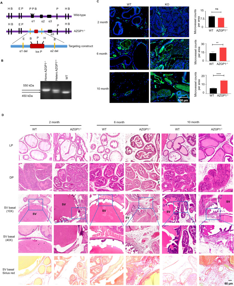
AZGP1 knockout and overexpressing prostate cancer cells were generated using a lentiviral system. The effects of AZGP1 under- or over-expression in prostate cancer cells were evaluated by in vitro cell proliferation, migration, and invasion assays. Heterozygous AZGP1 mice were obtained from European Mouse Mutant Archive (EMMA), and prostate tissues from homozygous knockout male mice were collected at 2, 6 and 10 months for histological analysis. In vivo xenografts generated from AZGP1 under- or over-expressing prostate cancer cells were used to determine the role of AZGP1 in prostate cancer tumor growth, and subsequent proteomics analysis was conducted to elucidate the mechanisms of AZGP1 action in prostate cancer progression. AZGP1 expression and microvessel density were measured in human prostate cancer samples on a tissue microarray of 215 independent patient samples.
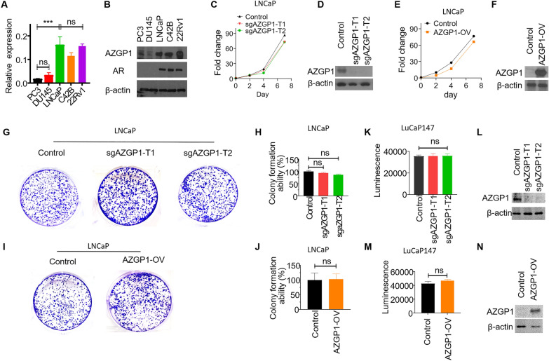
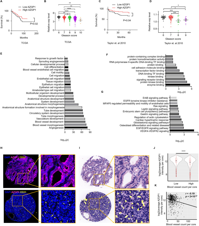
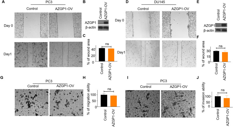
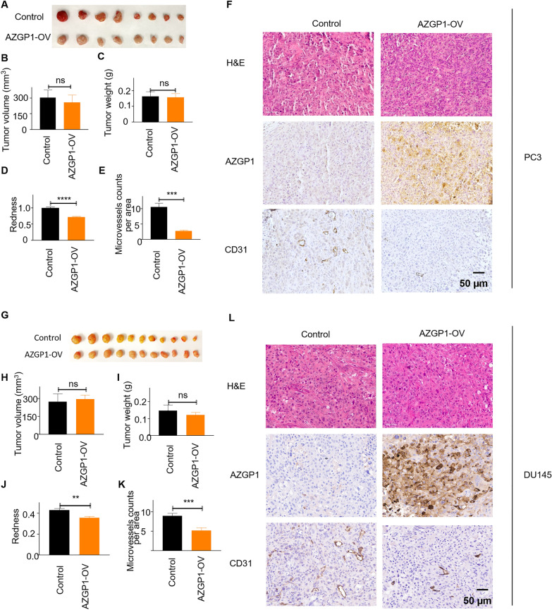
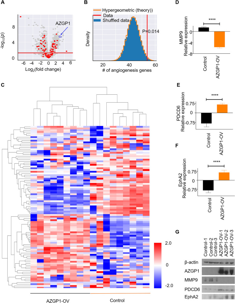
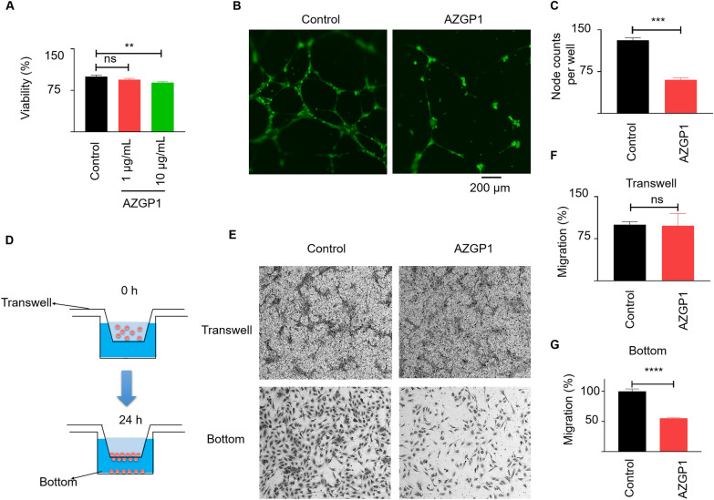
Neither the knockout nor overexpression of AZGP1 exhibited significant effects on prostate cancer cell proliferation, clonal growth, migration, or invasion in vitro. The prostates of AZGP1 mice initially appeared to have grossly normal morphology; however, we observed fibrosis in the periglandular stroma and higher blood vessel density in the mouse prostate by 6 months. In PC3 and DU145 mouse xenografts, over-expression of AZGP1 did not affect tumor growth. Instead, these tumors displayed decreased microvessel density compared to xenografts derived from PC3 and DU145 control cells, suggesting that AZGP1 functions to inhibit angiogenesis in prostate cancer. Proteomics profiling further indicated that, compared to control xenografts, AZGP1 overexpressing PC3 xenografts are enriched with angiogenesis pathway proteins, including YWHAZ, EPHA2, SERPINE1, and PDCD6, MMP9, GPX1, HSPB1, COL18A1, RNH1, and ANXA1. In vitro functional studies show that AZGP1 inhibits human umbilical vein endothelial cell proliferation, migration, tubular formation and branching. Additionally, tumor microarray analysis shows that AZGP1 expression is negatively correlated with blood vessel density in human prostate cancer tissues.
AZGP1 is a negative regulator of angiogenesis, such that loss of AZGP1 promotes angiogenesis in prostate cancer. AZGP1 likely exerts heterotypical effects on cells in the tumor microenvironment, such as stromal and endothelial cells. This study sheds light on the anti-angiogenic characteristics of AZGP1 in the prostate and provides a rationale to target AZGP1 to inhibit prostate cancer progression.
AZGP1表达缺失是一种与前列腺癌进展为去势抵抗、发生转移及疾病特异性生存不良相关的生物标志物。然而,有报道称前列腺癌中AZGP1高表达的细胞会增加增殖和侵袭能力。AZGP1在前列腺癌进展中的确切作用仍不清楚。
使用慢病毒系统构建AZGP1基因敲除和过表达的前列腺癌细胞。通过体外细胞增殖、迁移和侵袭实验评估AZGP1表达下调或上调对前列腺癌细胞的影响。从欧洲小鼠突变体库(EMMA)获得杂合子AZGP1小鼠,在2、6和10个月时收集纯合子基因敲除雄性小鼠的前列腺组织进行组织学分析。用AZGP1表达下调或上调的前列腺癌细胞构建体内异种移植模型,以确定AZGP1在前列腺癌肿瘤生长中的作用,并进行后续的蛋白质组学分析以阐明AZGP1在前列腺癌进展中的作用机制。在包含215个独立患者样本的组织微阵列上测量人前列腺癌样本中的AZGP1表达和微血管密度。
AZGP1的基因敲除和过表达对前列腺癌细胞的体外增殖、克隆生长、迁移或侵袭均无显著影响。AZGP1小鼠的前列腺最初外观形态基本正常;然而,到6个月时,我们观察到小鼠前列腺腺周基质纤维化且血管密度更高。在PC3和DU145小鼠异种移植模型中,AZGP1过表达不影响肿瘤生长。相反,与源自PC3和DU145对照细胞的异种移植瘤相比,这些肿瘤的微血管密度降低,这表明AZGP1在前列腺癌中发挥抑制血管生成的作用。蛋白质组学分析进一步表明,与对照异种移植瘤相比,过表达AZGP1的PC3异种移植瘤富含血管生成途径蛋白,包括YWHAZ、EPHA2、SERPINE1、PDCD6、MMP9、GPX1、HSPB1、COL18A1、RNH1和ANXA1。体外功能研究表明,AZGP1抑制人脐静脉内皮细胞的增殖、迁移、管状形成和分支。此外,肿瘤微阵列分析显示,AZGP1表达与人前列腺癌组织中的血管密度呈负相关。
AZGP1是血管生成的负调节因子,因此AZGP1缺失促进前列腺癌血管生成。AZGP1可能对肿瘤微环境中的细胞如基质细胞和内皮细胞发挥异型作用。本研究揭示了AZGP1在前列腺中的抗血管生成特性,并为靶向AZGP1以抑制前列腺癌进展提供了理论依据。