Clinics for Kidney and Hypertension Disease, Hannover Medical School, Hannover, Germany.
Mount Desert Island Biological Laboratory, Bar Harbor, Maine, USA.
Stem Cell Res Ther. 2024 May 3;15(1):132. doi: 10.1186/s13287-024-03739-8.
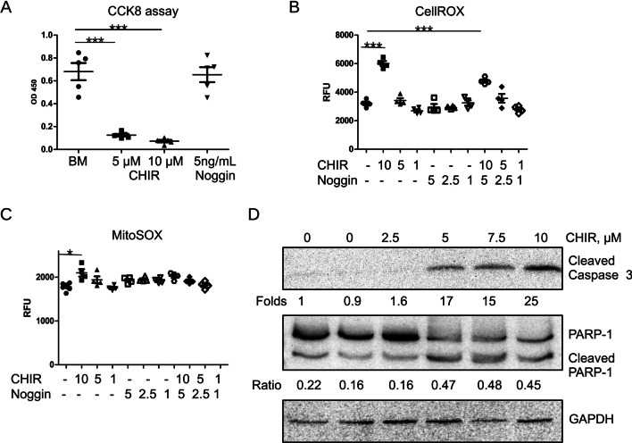
Induced pluripotent stem cells (iPSCs)-derived kidney organoids are a promising model for studying disease mechanisms and renal development. Despite several protocols having been developed, further improvements are needed to overcome existing limitations and enable a wider application of this model. One of the approaches to improve the differentiation of renal organoids in vitro is to include in the system cell types important for kidney organogenesis in vivo, such as macrophages. Another approach could be to improve cell survival. Mesodermal lineage differentiation is the common initial step of the reported protocols. The glycogen synthase kinase-3 (GSK-3) activity inhibitor, CHIR99021 (CHIR), is applied to induce mesodermal differentiation. It has been reported that CHIR simultaneously induces iPSCs apoptosis that can compromise cell differentiation. We thought to interfere with CHIR-induced apoptosis of iPSCs using rapamycin.
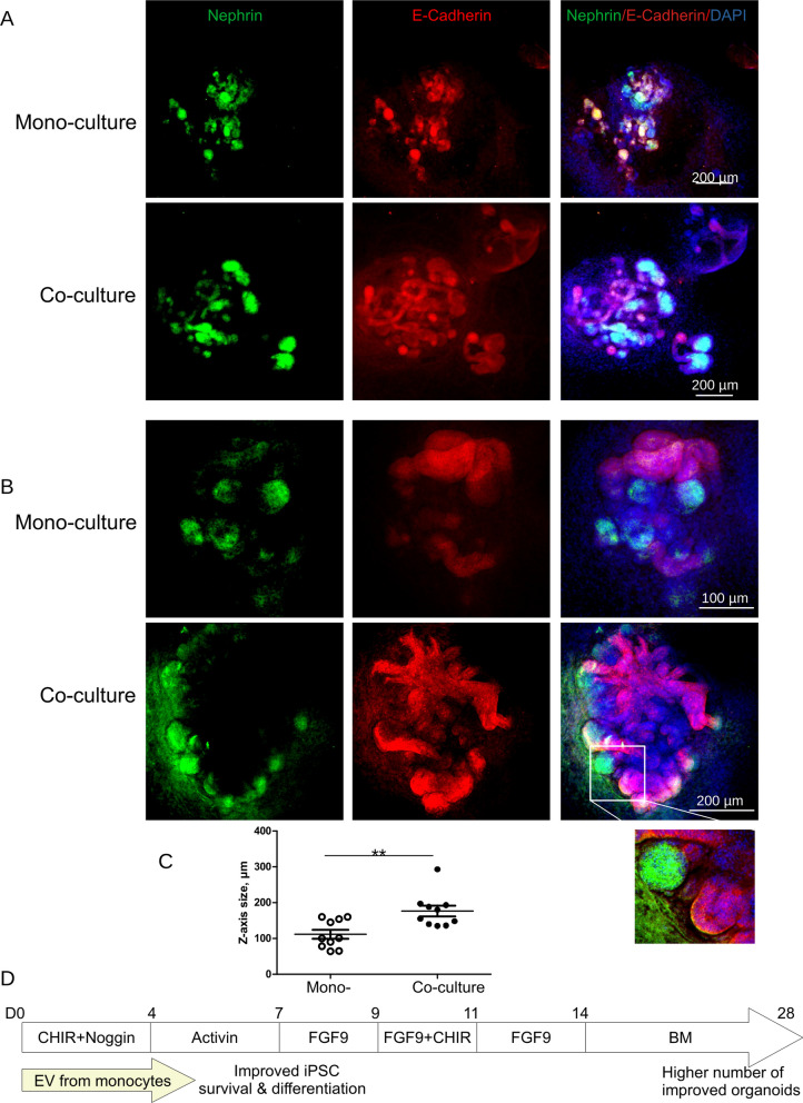
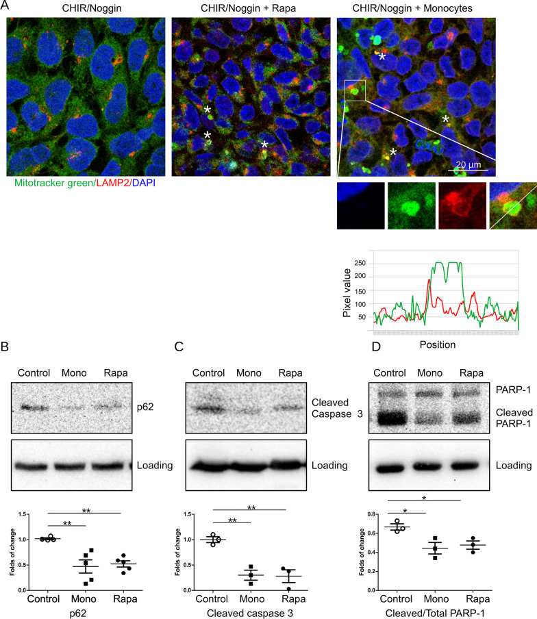
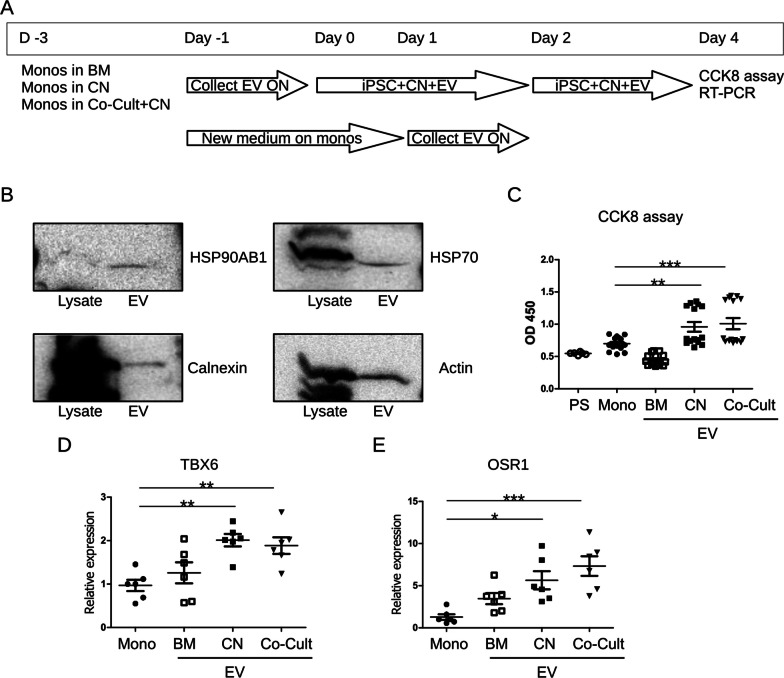
Differentiation of kidney organoids from human iPSCs was performed. Cell survival and autophagy were analyzed using Cell counting kit 8 (CCK8) kit and Autophagy detection kit. Cells were treated with rapamycin or co-cultured with human monocytes isolated from peripheral blood or iPSCs-macrophages using a transwell co-culture system. Monocyte-derived extracellular vesicles (EVs) were isolated using polyethylene glycol precipitation. Expression of apoptotic markers cleaved Caspase 3, Poly [ADP-ribose] polymerase 1 (PARP-1) and markers of differentiation T-Box Transcription Factor 6 (TBX6), odd-skipped related 1 (OSR1), Nephrin, E-Cadherin, Paired box gene 2 (Pax2) and GATA Binding Protein 3 (Gata3) was assessed by RT-PCR and western blotting. Organoids were imaged by 3D-confocal microscopy.
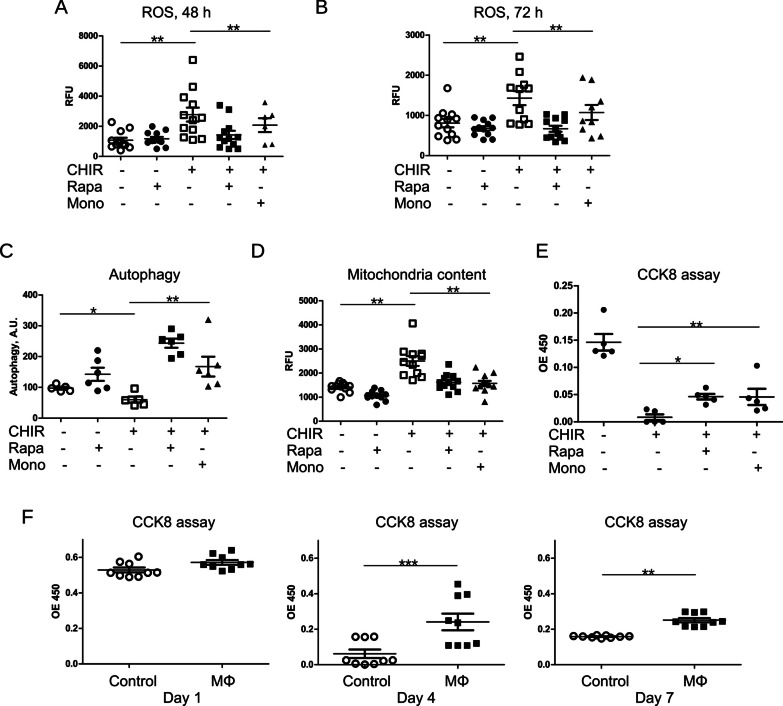
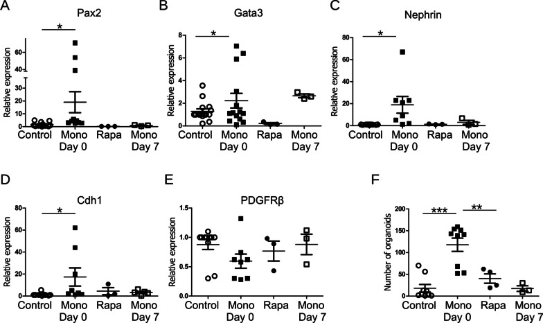
We observed that CHIR induced apoptosis of iPSCs during the initial stage of renal organoid differentiation. Underlying mechanisms implied the accumulation of reactive oxygen species and decreased autophagy. Activation of autophagy by rapamacin and by an indirect co-culture of differentiating iPSCs with iPSCs-macrophages and human peripheral blood monocytes prevented apoptosis induced by CHIR. Furthermore, monocytes (but not rapamycin) strongly promoted expression of renal differentiation markers and organoids development via released extracellular vesicles.
Our data suggest that co-culturing of iPSCs with human monocytes strongly improves differentiation of kidney organoids. An underlying mechanism of monocytic action implies, but not limited to, an increased autophagy in CHIR-treated iPSCs. Our findings enhance the utility of kidney organoid models.
诱导多能干细胞(iPSC)衍生的肾类器官是研究疾病机制和肾脏发育的有前途的模型。尽管已经开发了几种方案,但仍需要进一步改进,以克服现有局限性并更广泛地应用该模型。提高体外肾类器官分化的一种方法是在系统中加入体内肾发生中重要的细胞类型,如巨噬细胞。另一种方法可以是提高细胞存活率。中胚层谱系分化是报道的方案的共同初始步骤。糖原合酶激酶-3(GSK-3)活性抑制剂 CHIR99021(CHIR)用于诱导中胚层分化。据报道,CHIR 同时诱导 iPSC 细胞凋亡,这可能会影响细胞分化。我们考虑使用雷帕霉素来干扰 CHIR 诱导的 iPSC 细胞凋亡。
从人 iPSC 中分化肾类器官。使用细胞计数试剂盒 8(CCK8)试剂盒和自噬检测试剂盒分析细胞存活率和自噬。用雷帕霉素处理细胞或使用 Transwell 共培养系统与从外周血分离的人单核细胞或 iPSC-巨噬细胞共培养。使用聚乙二醇沉淀分离单核细胞衍生的细胞外囊泡(EVs)。通过 RT-PCR 和 Western blot 评估凋亡标志物 cleaved Caspase 3、多聚(ADP-核糖)聚合酶 1(PARP-1)和分化标志物 T-Box 转录因子 6(TBX6)、奇数跳过相关 1(OSR1)、nephrin、E-钙黏蛋白、配对盒基因 2(Pax2)和 GATA 结合蛋白 3(Gata3)的表达。通过 3D 共聚焦显微镜对类器官进行成像。
我们观察到 CHIR 在肾类器官分化的初始阶段诱导 iPSC 细胞凋亡。潜在机制暗示活性氧的积累和自噬减少。雷帕霉素和分化的 iPSC 与 iPSC-巨噬细胞和人外周血单核细胞的间接共培养激活自噬,可防止 CHIR 诱导的细胞凋亡。此外,单核细胞(而非雷帕霉素)通过释放细胞外囊泡强烈促进肾分化标志物的表达和类器官的发育。
我们的数据表明,iPSC 与人单核细胞的共培养可大大改善肾类器官的分化。单核细胞作用的潜在机制暗示,但不限于 CHIR 处理的 iPSC 中自噬增加。我们的发现增强了肾类器官模型的实用性。