From the Molecular Imaging Branch (Y.L., E.C.Y., M.J.B., S.A.H., T.E.P., K.M.M., N.S.L., P.L.C., B.T.), Center for Interventional Oncology (L.H., C.G., B.J.W.), Laboratory of Pathology (A.T., M.J.M.), and Urologic Oncology Branch (S.G., P.A.P.), National Cancer Institute, National Institutes of Health, 10 Center Dr, MSC 1182, Bldg 10, Rm B3B85, Bethesda, MD 20892; NVIDIA, Santa Clara, Calif (J.T., D.Y., Z.X., D.X.); Department of Radiology, Clinical Center, National Institutes of Health, Bethesda, Md (L.H., C.G., B.J.W.); and Department of Radiology, Singapore General Hospital, Singapore (Y.M.L.).
Radiology. 2024 May;311(2):e230750. doi: 10.1148/radiol.230750.
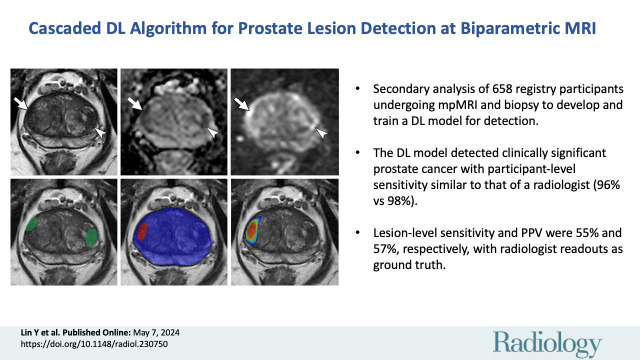
Background Multiparametric MRI (mpMRI) improves prostate cancer (PCa) detection compared with systematic biopsy, but its interpretation is prone to interreader variation, which results in performance inconsistency. Artificial intelligence (AI) models can assist in mpMRI interpretation, but large training data sets and extensive model testing are required. Purpose To evaluate a biparametric MRI AI algorithm for intraprostatic lesion detection and segmentation and to compare its performance with radiologist readings and biopsy results. Materials and Methods This secondary analysis of a prospective registry included consecutive patients with suspected or known PCa who underwent mpMRI, US-guided systematic biopsy, or combined systematic and MRI/US fusion-guided biopsy between April 2019 and September 2022. All lesions were prospectively evaluated using Prostate Imaging Reporting and Data System version 2.1. The lesion- and participant-level performance of a previously developed cascaded deep learning algorithm was compared with histopathologic outcomes and radiologist readings using sensitivity, positive predictive value (PPV), and Dice similarity coefficient (DSC). Results A total of 658 male participants (median age, 67 years [IQR, 61-71 years]) with 1029 MRI-visible lesions were included. At histopathologic analysis, 45% (294 of 658) of participants had lesions of International Society of Urological Pathology (ISUP) grade group (GG) 2 or higher. The algorithm identified 96% (282 of 294; 95% CI: 94%, 98%) of all participants with clinically significant PCa, whereas the radiologist identified 98% (287 of 294; 95% CI: 96%, 99%; = .23). The algorithm identified 84% (103 of 122), 96% (152 of 159), 96% (47 of 49), 95% (38 of 40), and 98% (45 of 46) of participants with ISUP GG 1, 2, 3, 4, and 5 lesions, respectively. In the lesion-level analysis using radiologist ground truth, the detection sensitivity was 55% (569 of 1029; 95% CI: 52%, 58%), and the PPV was 57% (535 of 934; 95% CI: 54%, 61%). The mean number of false-positive lesions per participant was 0.61 (range, 0-3). The lesion segmentation DSC was 0.29. Conclusion The AI algorithm detected cancer-suspicious lesions on biparametric MRI scans with a performance comparable to that of an experienced radiologist. Moreover, the algorithm reliably predicted clinically significant lesions at histopathologic examination. ClinicalTrials.gov Identifier: NCT03354416 © RSNA, 2024
背景 与系统活检相比,多参数 MRI(mpMRI)可提高前列腺癌(PCa)的检出率,但其解读容易受到读者间差异的影响,从而导致性能不一致。人工智能(AI)模型可以辅助 mpMRI 解读,但需要大量的训练数据集和广泛的模型测试。目的 评估一种双参数 MRI AI 算法在前列腺内病变检测和分割中的性能,并比较其与放射科医生阅读和活检结果的性能。材料与方法 本研究对 2019 年 4 月至 2022 年 9 月间连续接受 mpMRI、超声引导下系统活检或系统和 MRI/US 融合引导下联合活检的疑似或已知前列腺癌患者的前瞻性登记进行了二次分析。所有病变均采用前列腺影像报告和数据系统第 2.1 版进行前瞻性评估。使用灵敏度、阳性预测值(PPV)和 Dice 相似系数(DSC)比较了先前开发的级联深度学习算法在病变和参与者水平上的表现与组织病理学结果和放射科医生的阅读结果。结果 共纳入 658 名男性参与者(中位年龄,67 岁[IQR,61-71 岁])和 1029 个 MRI 可见病变。在组织病理学分析中,45%(294/658)的参与者存在国际泌尿病理学会(ISUP)分级组(GG)2 级或更高的病变。该算法可识别出 96%(282/294;95%CI:94%,98%)的所有有临床意义的 PCa 患者,而放射科医生识别出 98%(287/294;95%CI:96%,99%; =.23)。该算法分别识别出 122 名、159 名、49 名、40 名和 46 名参与者的 ISUP GG 1、2、3、4 和 5 级病变,其识别率分别为 84%(103/122)、96%(152/159)、96%(47/49)、95%(38/40)和 98%(45/46)。在使用放射科医生地面实况的病变水平分析中,检测灵敏度为 55%(569/1029;95%CI:52%,58%),PPV 为 57%(535/934;95%CI:54%,61%)。每个参与者的平均假阳性病变数为 0.61(范围,0-3)。病变分割的 DSC 为 0.29。结论 AI 算法可在双参数 MRI 扫描中检测可疑癌性病变,其性能与经验丰富的放射科医生相当。此外,该算法还可可靠地预测组织病理学检查中有临床意义的病变。ClinicalTrials.gov 标识符:NCT03354416 ©RSNA,2024