Underwood S R, Walton S, Laming P J, Jarritt P H, Ell P J, Emanuel R W, Swanton R H
Br Heart J. 1985 Feb;53(2):216-22. doi: 10.1136/hrt.53.2.216.
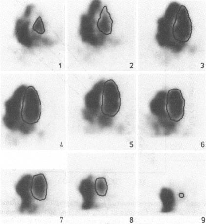
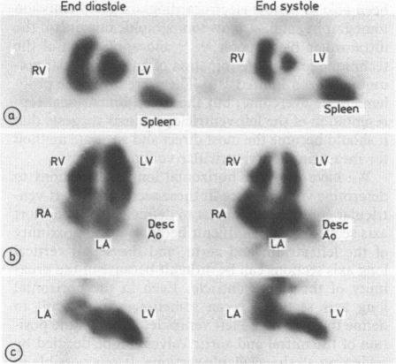
Electrocardiogram gated single photon emission computed tomography of the intracardiac blood pools is a recent development that involves the acquisition of images in multiple projections after in vivo erythrocyte labelling with technetium-99m and reconstruction of these images into tomographic sections in any desired plane. The technique was used in 25 subjects to measure left ventricular volume, by summing the areas of the ventricle in each of the tomographic sections, and the results compared with those using a counts based (non-geometric) technique from planar radionuclide ventriculography. Endocardium was defined with the aid of a contour at 43% of maximum left ventricular counts, and this contour was validated for a left ventricular phantom. Correlation between tomographic and counts based left ventricular volume was close. Similarly, ejection fraction correlated well. The technique is therefore an accurate method for determining left ventricular volume and ejection fraction, avoiding the assumptions about shape made by other geometric methods.
心电门控心脏血池单光子发射计算机断层扫描是一项最新技术,它涉及在用99m锝对体内红细胞进行标记后,从多个投影方向采集图像,并将这些图像重建为任意所需平面的断层切片。该技术应用于25名受试者,通过累加每个断层切片中心室的面积来测量左心室容积,并将结果与使用平面放射性核素心室造影的基于计数(非几何)技术所得结果进行比较。借助左心室最大计数43%处的轮廓来定义心内膜,并且该轮廓在左心室模型上得到了验证。断层扫描法与基于计数法测得的左心室容积之间相关性密切。同样,射血分数相关性良好。因此,该技术是一种确定左心室容积和射血分数的准确方法,避免了其他几何方法对形状所做的假设。