Underwood S R, Rees R S, Savage P E, Klipstein R H, Firmin D N, Fox K M, Poole-Wilson P A, Longmore D B
Br Heart J. 1986 Oct;56(4):334-40. doi: 10.1136/hrt.56.4.334.
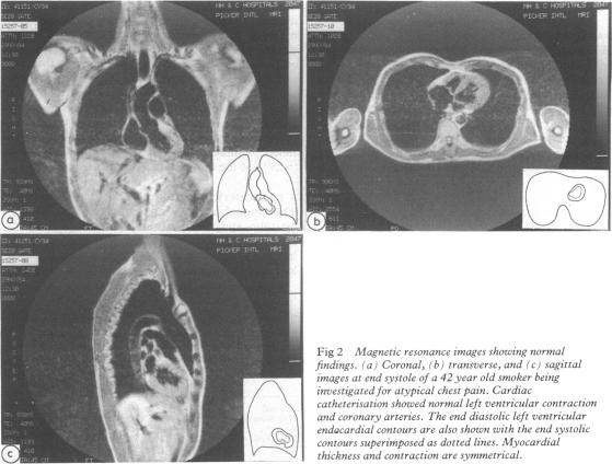
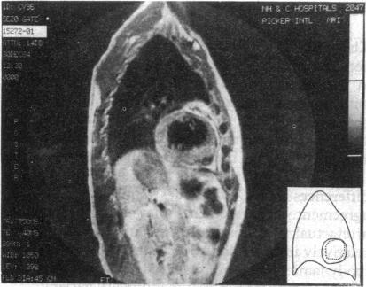
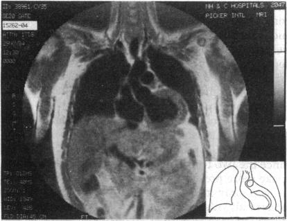
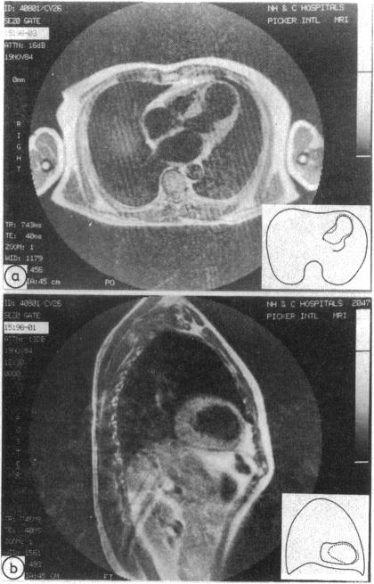
The ability of magnetic resonance to determine regional left ventricular function was investigated in 18 patients--13 with coronary artery disease (nine with previous infarction), one with congestive cardiomyopathy, one with mitral stenosis, one with an atrial septal defect, and two without detectable cardiac abnormality. Coronal magnetic resonance images were acquired through the aortic valve and sagittal images were acquired in the plane of widest diameter of the left ventricle seen in the coronal image, both at end diastole and end systole. Regional wall motion assessed by magnetic resonance was compared with the results of anteroposterior and left lateral x ray ventriculograms by two independent observers. The left ventricular wall was divided into three segments in each plane and the motion of the segments was classified as normal, hypokinetic, akinetic, or dyskinetic. Muscle thickness was measured in each segment of the magnetic resonance images and was considered to be abnormal if in the systolic images it was less than 75% of that in neighbouring segments or if it failed to increase by at least 25% between diastole and systole. Wall motion assessments by the two methods agreed in 68 of 105 segments analysed, but differed by one class in 32 segments and by two classes in five segments. The differences can be explained by the conditions under which the investigations were performed and by the disparity between a tomographic section and an x ray projection. Magnetic resonance showed 25 segments to have abnormal wall thickness. Only one patient with infarction did not have an area of wall thinning and no patient without infarction had an area of thinning. It is concluded that magnetic resonance allows an accurate non-invasive assessment of left ventricular wall motion and thickness.
对18例患者进行了研究,以探讨磁共振测定局部左心室功能的能力。其中13例患有冠状动脉疾病(9例有陈旧性心肌梗死),1例患有充血性心肌病,1例患有二尖瓣狭窄,1例患有房间隔缺损,2例未检测到心脏异常。在舒张末期和收缩末期,通过主动脉瓣获取冠状面磁共振图像,并在冠状面图像中左心室最大直径平面获取矢状面图像。由两名独立观察者将磁共振评估的局部壁运动与前后位和左侧位X线心室造影结果进行比较。在每个平面将左心室壁分为三个节段,并将节段的运动分为正常、运动减弱、无运动或运动障碍。在磁共振图像的每个节段测量肌肉厚度,如果在收缩期图像中该厚度小于相邻节段的75%,或者在舒张期和收缩期之间未能至少增加25%,则认为该厚度异常。在分析的105个节段中,两种方法对壁运动的评估在68个节段中一致,但在32个节段中相差一级,在5个节段中相差两级。这些差异可以通过检查所采用的条件以及断层切片与X线投影之间的差异来解释。磁共振显示25个节段的壁厚度异常。只有1例梗死患者没有壁变薄区域,没有梗死的患者也没有变薄区域。结论是,磁共振能够对左心室壁运动和厚度进行准确的非侵入性评估。