Huaman Celeste, Clouse Caitlyn, Rader Madeline, Yan Lianying, Bai Shuangyi, Gunn Bronwyn M, Amaya Moushimi, Laing Eric D, Broder Christopher C, Schaefer Brian C
Department of Microbiology and Immunology, Uniformed Services University, Bethesda, MD, USA.
Henry M. Jackson Foundation for the Advancement of Military Medicine, Inc., Rockville, MD, USA.
Front Chem Biol. 2024;3. doi: 10.3389/fchbi.2024.1363498. Epub 2024 Mar 18.
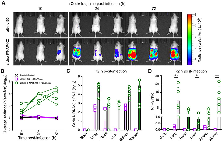
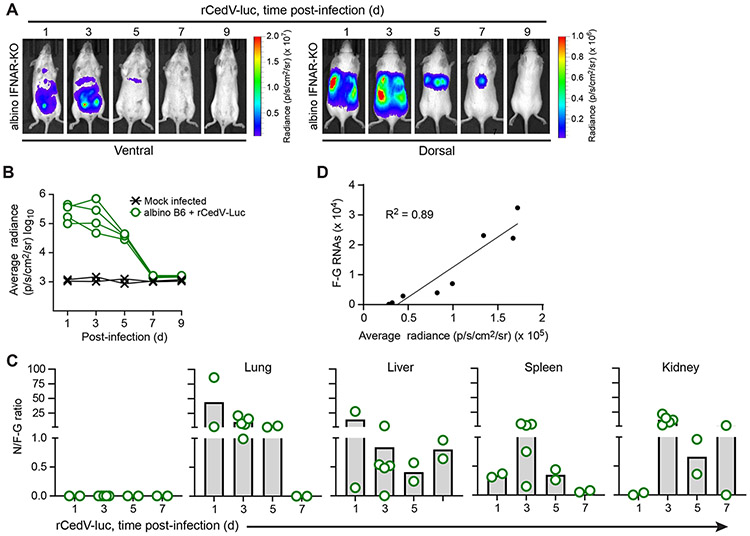
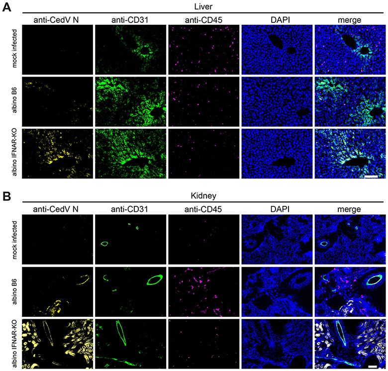
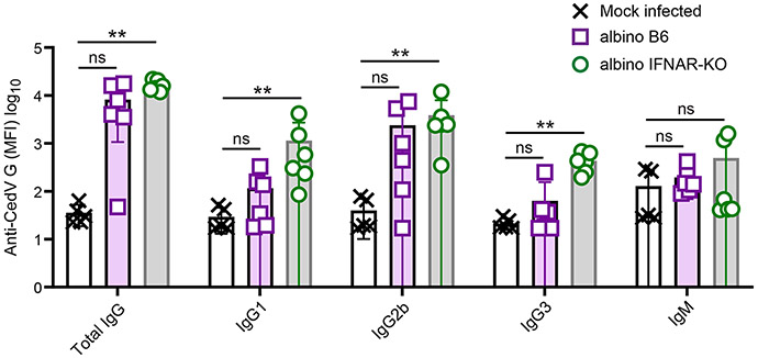
Henipaviruses are enveloped single-stranded, negative-sense RNA viruses of the paramyxovirus family. Two henipaviruses, Nipah virus and Hendra virus, cause a systemic respiratory and/or neurological disease in humans and ten additional species of mammals, with a high fatality rate. Because of their highly pathogenic nature, Nipah virus and Hendra virus are categorized as BSL-4 pathogens, which limits the number and scope of translational research studies on these important human pathogens. To begin to address this limitation, we are developing a BSL-2 model of authentic henipavirus infection in mice, using the non-pathogenic henipavirus, Cedar virus. Notably, wild-type mice are highly resistant to Hendra virus and Nipah virus infection. However, previous work has shown that mice lacking expression of the type I interferon receptor (IFNAR-KO mice) are susceptible to both viruses. Here, we show that luciferase-expressing recombinant Cedar virus (rCedV-luc) is also able to replicate and establish a transient infection in IFNAR-KO mice, but not in wild-type mice. Using longitudinal bioluminescence imaging (BLI) of luciferase expression, we detected rCedV-luc replication as early as 10 h post-infection. Viral replication peaks between days 1 and 3 post-infection, and declines to levels undetectable by BLI by 7 days post-infection. Immunohistochemistry is consistent with viral infection and replication in endothelial cells and other non-immune cell types within tissue parenchyma. Serology analyses demonstrate significant IgG responses to the Cedar virus surface glycoprotein with potent neutralizing activity in IFNAR-KO mice, whereas antibody responses in wild-type animals were non-significant. Overall, these data suggest that rCedV-luc infection of IFNAR-KO mice represents a viable platform for the study of henipavirus replication, anti-henipavirus host responses and henipavirus-directed therapeutics.
亨尼帕病毒是副粘病毒科的包膜单链负义RNA病毒。两种亨尼帕病毒,即尼帕病毒和亨德拉病毒,可在人类和另外10种哺乳动物中引起全身性呼吸道疾病和/或神经系统疾病,致死率很高。由于其高致病性,尼帕病毒和亨德拉病毒被归类为生物安全4级病原体,这限制了对这些重要人类病原体进行转化研究的数量和范围。为了开始解决这一限制,我们正在利用非致病性亨尼帕病毒——雪松病毒,开发一种在小鼠中进行真实亨尼帕病毒感染的生物安全2级模型。值得注意的是,野生型小鼠对亨德拉病毒和尼帕病毒感染具有高度抗性。然而,先前的研究表明,缺乏I型干扰素受体表达的小鼠(I型干扰素受体基因敲除小鼠)对这两种病毒敏感。在这里,我们表明,表达荧光素酶的重组雪松病毒(rCedV-luc)也能够在I型干扰素受体基因敲除小鼠中复制并建立短暂感染,但在野生型小鼠中则不能。通过对荧光素酶表达进行纵向生物发光成像(BLI),我们在感染后10小时就检测到了rCedV-luc的复制。病毒复制在感染后第1天至第3天达到峰值,并在感染后7天下降至BLI检测不到的水平。免疫组织化学结果与组织实质内内皮细胞和其他非免疫细胞类型中的病毒感染和复制情况一致。血清学分析表明,I型干扰素受体基因敲除小鼠对雪松病毒表面糖蛋白有显著的IgG反应,并具有强大的中和活性,而野生型动物的抗体反应不显著。总体而言,这些数据表明,I型干扰素受体基因敲除小鼠的rCedV-luc感染代表了一个可行的平台,可用于研究亨尼帕病毒复制、抗亨尼帕病毒宿主反应以及针对亨尼帕病毒的治疗方法。