• 文献检索
  • 文档翻译
  • 深度研究
  • 学术资讯
  • Suppr Zotero 插件Zotero 插件
  • 邀请有礼
  • 套餐&价格
  • 历史记录
应用&插件
Suppr Zotero 插件Zotero 插件浏览器插件Mac 客户端Windows 客户端微信小程序
定价
高级版会员购买积分包购买API积分包
服务
文献检索文档翻译深度研究API 文档MCP 服务
关于我们
关于 Suppr公司介绍联系我们用户协议隐私条款
关注我们

Suppr 超能文献

核心技术专利:CN118964589B侵权必究
粤ICP备2023148730 号-1Suppr @ 2026

文献检索

告别复杂PubMed语法,用中文像聊天一样搜索,搜遍4000万医学文献。AI智能推荐,让科研检索更轻松。

立即免费搜索

文件翻译

保留排版,准确专业,支持PDF/Word/PPT等文件格式,支持 12+语言互译。

免费翻译文档

深度研究

AI帮你快速写综述,25分钟生成高质量综述,智能提取关键信息,辅助科研写作。

立即免费体验

实验性百草枯中毒后肺结构重塑模式。肺泡内纤维化的形态发生。

Patterns of pulmonary structural remodeling after experimental paraquat toxicity. The morphogenesis of intraalveolar fibrosis.

作者信息

Fukuda Y, Ferrans V J, Schoenberger C I, Rennard S I, Crystal R G

出版信息

Am J Pathol. 1985 Mar;118(3):452-75.

PMID:3883797
原文链接:https://pmc.ncbi.nlm.nih.gov/articles/PMC1887950/
Abstract

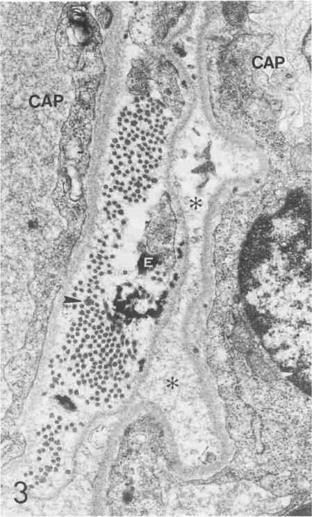
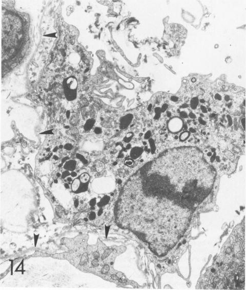
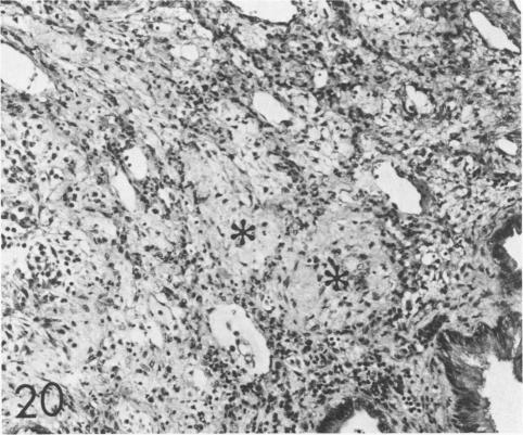
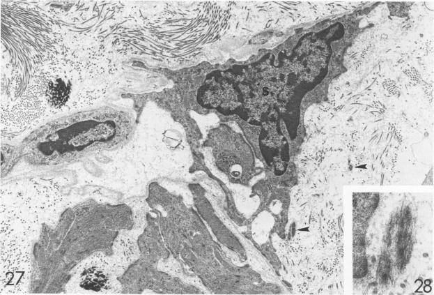
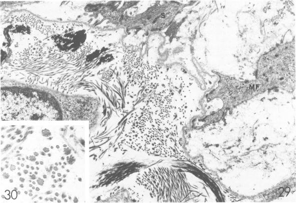
For a study of the evolution of interstitial and intraalveolar fibrosis, ultrastructural and immunohistochemical observations were made of the lungs of 16 cynomolgous monkeys given 1 or 2 injections of 10 mg/kg of paraquat and sacrificed 2 days to 8 weeks later. At 2-3 days, alveolar epithelial cells were denuded in many areas, and fibronectin was conspicuous in alveolar spaces. At 1 week, fibroblasts and inflammatory cells were migrating through gaps in the denuded epithelial basement membranes; Type II cells were regenerating in some areas. At 3-4 weeks, alveoli developing intraalveolar fibrosis contained many myofibroblasts, collagen fibrils, and small elastic fibers; fibrotic alveolar walls were lined by metaplastic squamous cells and bronchiolar epithelial cells. Spiraling collagen fibrils were found in interstitium but not in alveolar spaces, which suggests that they were formed from breakdown of collagen. Newly formed intraalveolar collagen was mainly Type I. At 8 weeks, intraalveolar fibrosis had led to extensive remodeling, with new glandlike alveoli lined by Type II cells; alveoli without intraalveolar fibrosis had more normal architecture. Thus, intraalveolar fibrosis in paraquattreated lung is mediated by intraalveolar migration of interstitial cells, through gaps in the epithelial basement membranes, after epithelial injury. This is followed by connective tissue synthesis on the luminal side of the epithelial basement membrane, by differentiation of interstitial cells into myofibroblasts and smooth-muscle cells, by incorporation of areas of intraalveolar fibrosis into the interstitium, and by coalescence of alveolar walls. Intraalveolar fibrosis is more important than interstitial fibrosis in the structural remodeling that occurs in paraquattreated lung, because it results in obliteration of alveoli, coalescence of alveolar walls, and loss of functional alveolar-capillary units.

摘要

为研究间质性和肺泡内纤维化的演变,对16只食蟹猴的肺进行了超微结构和免疫组织化学观察。这些食蟹猴接受了1或2次10mg/kg百草枯注射,并于2天至8周后处死。在2 - 3天时,许多区域的肺泡上皮细胞剥脱,肺泡腔内纤连蛋白明显。在1周时,成纤维细胞和炎性细胞通过剥脱的上皮基底膜间隙迁移;部分区域Ⅱ型细胞再生。在3 - 4周时,发生肺泡内纤维化的肺泡含有许多肌成纤维细胞、胶原纤维和小弹性纤维;纤维化的肺泡壁由化生的鳞状细胞和细支气管上皮细胞衬里。在间质中发现螺旋状胶原纤维,但肺泡腔内未发现,这表明它们是由胶原分解形成的。新形成的肺泡内胶原主要为Ⅰ型。在8周时,肺泡内纤维化导致广泛重塑,新的腺泡样肺泡由Ⅱ型细胞衬里;无肺泡内纤维化的肺泡结构更正常。因此,百草枯处理的肺中肺泡内纤维化是由间质细胞在肺泡上皮损伤后通过上皮基底膜间隙向肺泡内迁移介导的。随后,在上皮基底膜腔面进行结缔组织合成,间质细胞分化为肌成纤维细胞和平滑肌细胞,肺泡内纤维化区域并入间质,肺泡壁融合。在百草枯处理的肺发生的结构重塑中,肺泡内纤维化比间质性纤维化更重要,因为它导致肺泡闭塞、肺泡壁融合以及功能性肺泡 - 毛细血管单位丧失。

https://cdn.ncbi.nlm.nih.gov/pmc/blobs/51cc/1887950/dca984458b5a/amjpathol00174-0136-b.jpg
https://cdn.ncbi.nlm.nih.gov/pmc/blobs/51cc/1887950/558c87b419cd/amjpathol00174-0119-a.jpg
https://cdn.ncbi.nlm.nih.gov/pmc/blobs/51cc/1887950/504ffe7504c0/amjpathol00174-0119-b.jpg
https://cdn.ncbi.nlm.nih.gov/pmc/blobs/51cc/1887950/5587495977d1/amjpathol00174-0119-c.jpg
https://cdn.ncbi.nlm.nih.gov/pmc/blobs/51cc/1887950/a8b5c9f5e933/amjpathol00174-0121-a.jpg
https://cdn.ncbi.nlm.nih.gov/pmc/blobs/51cc/1887950/4c79e1449a6b/amjpathol00174-0121-b.jpg
https://cdn.ncbi.nlm.nih.gov/pmc/blobs/51cc/1887950/4abd76a80229/amjpathol00174-0121-c.jpg
https://cdn.ncbi.nlm.nih.gov/pmc/blobs/51cc/1887950/acb8bca9d841/amjpathol00174-0121-d.jpg
https://cdn.ncbi.nlm.nih.gov/pmc/blobs/51cc/1887950/17f9528dc312/amjpathol00174-0121-e.jpg
https://cdn.ncbi.nlm.nih.gov/pmc/blobs/51cc/1887950/e8ae6ab5f845/amjpathol00174-0121-f.jpg
https://cdn.ncbi.nlm.nih.gov/pmc/blobs/51cc/1887950/87a56ebe1843/amjpathol00174-0123-a.jpg
https://cdn.ncbi.nlm.nih.gov/pmc/blobs/51cc/1887950/8f74d99c6995/amjpathol00174-0123-b.jpg
https://cdn.ncbi.nlm.nih.gov/pmc/blobs/51cc/1887950/5a096249fbb1/amjpathol00174-0125-a.jpg
https://cdn.ncbi.nlm.nih.gov/pmc/blobs/51cc/1887950/0e8db4c6e661/amjpathol00174-0125-b.jpg
https://cdn.ncbi.nlm.nih.gov/pmc/blobs/51cc/1887950/e9436446016d/amjpathol00174-0125-c.jpg
https://cdn.ncbi.nlm.nih.gov/pmc/blobs/51cc/1887950/a52c78d0b287/amjpathol00174-0126-a.jpg
https://cdn.ncbi.nlm.nih.gov/pmc/blobs/51cc/1887950/28f4ec5507f7/amjpathol00174-0126-b.jpg
https://cdn.ncbi.nlm.nih.gov/pmc/blobs/51cc/1887950/ff351790a6d2/amjpathol00174-0127-a.jpg
https://cdn.ncbi.nlm.nih.gov/pmc/blobs/51cc/1887950/edf98fa8bada/amjpathol00174-0127-b.jpg
https://cdn.ncbi.nlm.nih.gov/pmc/blobs/51cc/1887950/e5c04d7f6e17/amjpathol00174-0127-c.jpg
https://cdn.ncbi.nlm.nih.gov/pmc/blobs/51cc/1887950/c0b06e6d3bac/amjpathol00174-0127-d.jpg
https://cdn.ncbi.nlm.nih.gov/pmc/blobs/51cc/1887950/f2acb2dbc611/amjpathol00174-0127-e.jpg
https://cdn.ncbi.nlm.nih.gov/pmc/blobs/51cc/1887950/dd9bab978d8b/amjpathol00174-0127-f.jpg
https://cdn.ncbi.nlm.nih.gov/pmc/blobs/51cc/1887950/c2e0a47b61b5/amjpathol00174-0128-a.jpg
https://cdn.ncbi.nlm.nih.gov/pmc/blobs/51cc/1887950/d78dec74b4ed/amjpathol00174-0130-a.jpg
https://cdn.ncbi.nlm.nih.gov/pmc/blobs/51cc/1887950/ec46f4e0eb3a/amjpathol00174-0130-b.jpg
https://cdn.ncbi.nlm.nih.gov/pmc/blobs/51cc/1887950/07bcc3cde924/amjpathol00174-0132-a.jpg
https://cdn.ncbi.nlm.nih.gov/pmc/blobs/51cc/1887950/a3ef7e08cce1/amjpathol00174-0132-b.jpg
https://cdn.ncbi.nlm.nih.gov/pmc/blobs/51cc/1887950/47d7ed6dfba1/amjpathol00174-0134-a.jpg
https://cdn.ncbi.nlm.nih.gov/pmc/blobs/51cc/1887950/760c353fb177/amjpathol00174-0134-b.jpg
https://cdn.ncbi.nlm.nih.gov/pmc/blobs/51cc/1887950/2a955ecdf790/amjpathol00174-0134-c.jpg
https://cdn.ncbi.nlm.nih.gov/pmc/blobs/51cc/1887950/d01edc753a7e/amjpathol00174-0134-d.jpg
https://cdn.ncbi.nlm.nih.gov/pmc/blobs/51cc/1887950/c90856e1ada5/amjpathol00174-0134-e.jpg
https://cdn.ncbi.nlm.nih.gov/pmc/blobs/51cc/1887950/bc6336928db6/amjpathol00174-0136-a.jpg
https://cdn.ncbi.nlm.nih.gov/pmc/blobs/51cc/1887950/dca984458b5a/amjpathol00174-0136-b.jpg

相似文献

1
Patterns of pulmonary structural remodeling after experimental paraquat toxicity. The morphogenesis of intraalveolar fibrosis.实验性百草枯中毒后肺结构重塑模式。肺泡内纤维化的形态发生。
Am J Pathol. 1985 Mar;118(3):452-75.
2
The role of intraalveolar fibrosis in the process of pulmonary structural remodeling in patients with diffuse alveolar damage.肺泡内纤维化在弥漫性肺泡损伤患者肺结构重塑过程中的作用。
Am J Pathol. 1987 Jan;126(1):171-82.
3
Pulmonary elastic fiber degradation in paraquat toxicity. An electron microscopic immunohistochemical study.百草枯中毒时肺弹性纤维降解。一项电子显微镜免疫组织化学研究。
J Submicrosc Cytol Pathol. 1988 Jan;20(1):15-23.
4
Evolution of metaplastic squamous cells of alveolar walls in pulmonary fibrosis produced by paraquat. An ultrastructural and immunohistochemical study.百草枯所致肺纤维化中肺泡壁化生鳞状细胞的演变。一项超微结构和免疫组织化学研究。
Virchows Arch B Cell Pathol Incl Mol Pathol. 1989;58(1):27-43. doi: 10.1007/BF02890057.
5
[Development of connective tissue and surfactant production in fetal lung].[胎儿肺脏中结缔组织的发育及表面活性物质的产生]
Nihon Kyobu Shikkan Gakkai Zasshi. 1991 Jan;29(1):24-8.
6
Structure of alveolar epithelial cells in patients with fibrotic lung disorders.纤维化肺部疾病患者肺泡上皮细胞的结构
Lab Invest. 1982 Jan;46(1):39-53.
7
Consequences of prolonged inhalation of ozone on F344/N rats: collaborative studies. Part VIII: Morphometric analysis of structural alterations in alveolar regions.长期吸入臭氧对F344/N大鼠的影响:合作研究。第八部分:肺泡区域结构改变的形态计量分析。
Res Rep Health Eff Inst. 1995 Mar(65 Pt 8-9):3-39; discussion 99-110.
8
Intraluminal fibrosis and elastic fiber degradation lead to lung remodeling in pulmonary Langerhans cell granulomatosis (histiocytosis X).管腔内纤维化和弹性纤维降解导致肺朗格汉斯细胞组织细胞增生症(组织细胞增多症X)中的肺部重塑。
Am J Pathol. 1990 Aug;137(2):415-24.
9
Anchoring fibrils. A new connective tissue structure in fibrotic lung disease.锚定原纤维。纤维化肺病中的一种新的结缔组织结构。
Am J Pathol. 1978 Aug;92(2):389-410.
10
Bronchiolarized metaplasia and interstitial fibrosis in rat lungs chronically exposed to high ambient levels of ozone.长期暴露于高环境水平臭氧的大鼠肺部出现细支气管化生和间质纤维化。
Toxicol Appl Pharmacol. 1995 Oct;134(2):251-63. doi: 10.1006/taap.1995.1191.

引用本文的文献

1
The Hidden Pandemic of COVID-19-Induced Organizing Pneumonia.新冠病毒诱发的机化性肺炎这一隐匿的大流行疾病
Pharmaceuticals (Basel). 2022 Dec 16;15(12):1574. doi: 10.3390/ph15121574.
2
Thoracic radiographic features of fatal paraquat intoxication in eleven dogs.11 例犬致死性百草枯中毒的胸部 X 线特征。
Vet Q. 2021 Dec;41(1):217-225. doi: 10.1080/01652176.2021.1950945.
3
Glucocorticoid with cyclophosphamide for oral paraquat poisoning.糖皮质激素联合环磷酰胺治疗口服百草枯中毒。

本文引用的文献

1
Laminin-secreting yolk sac carcinoma of the rat. Biochemical and electron immunohistochemical studies.大鼠层粘连蛋白分泌性卵黄囊癌。生化及电子免疫组织化学研究。
Lab Invest. 1982 Sep;47(3):247-57.
2
[Paraquat intoxication, an ultra-structural study of lung lesions. In connection with a case study (author's transl)].百草枯中毒:肺损伤的超微结构研究。结合病例分析(作者译)
Arch Anat Cytol Pathol. 1982;30(1):33-8.
3
Long-term morphologic and biochemical features of experimentally induced lung fibrosis in the mouse.小鼠实验性诱导肺纤维化的长期形态学和生化特征。
Cochrane Database Syst Rev. 2021 Jun 30;6(6):CD008084. doi: 10.1002/14651858.CD008084.pub5.
4
CD34+ derived macrophage and dendritic cells display differential responses to paraquat.CD34+ 衍生的巨噬细胞和树突状细胞对百草枯表现出不同的反应。
Toxicol In Vitro. 2021 Sep;75:105198. doi: 10.1016/j.tiv.2021.105198. Epub 2021 Jun 9.
5
Alveolar Epithelial Denudation Is a Major Factor in the Pathogenesis of Pleuroparenchymal Fibroelastosis.肺泡上皮剥脱是胸膜实质纤维弹性组织增生症发病机制中的一个主要因素。
J Clin Med. 2021 Feb 24;10(5):895. doi: 10.3390/jcm10050895.
6
Osteonecrosis of Femoral Head, An Overlooked Long-Term Complication after Paraquat Intoxication: A Retrospective Cohort Study.股骨头坏死,百草枯中毒后被忽视的长期并发症:一项回顾性队列研究。
Sci Rep. 2020 Jun 1;10(1):8827. doi: 10.1038/s41598-020-65756-w.
7
The role of club cell phenoconversion and migration in idiopathic pulmonary fibrosis.克拉拉细胞表型转化和迁移在特发性肺纤维化中的作用。
Aging (Albany NY). 2016 Nov 29;8(11):3091-3109. doi: 10.18632/aging.101115.
8
Curcumin inhibits paraquat induced lung inflammation and fibrosis by extracellular matrix modifications in mouse model.姜黄素通过改变细胞外基质抑制百草枯诱导的小鼠肺部炎症和纤维化。
Inflammopharmacology. 2016 Dec;24(6):335-345. doi: 10.1007/s10787-016-0286-z. Epub 2016 Oct 20.
9
Pulmonary fibrosis: patterns and perpetrators.肺纤维化:模式与元凶。
J Clin Invest. 2012 Aug;122(8):2756-62. doi: 10.1172/JCI60323. Epub 2012 Aug 1.
10
Organizing pneumonia: a kaleidoscope of concepts and morphologies.机化性肺炎:概念与形态的万花筒。
Eur Radiol. 2011 Nov;21(11):2244-54. doi: 10.1007/s00330-011-2191-6. Epub 2011 Jul 10.
Lab Invest. 1982 Apr;46(4):438-49.
4
Studies in cutaneous aging: I. The elastic fiber network.皮肤衰老的研究:I. 弹性纤维网络。
J Invest Dermatol. 1982 May;78(5):434-43. doi: 10.1111/1523-1747.ep12507866.
5
Structure of alveolar epithelial cells in patients with fibrotic lung disorders.纤维化肺部疾病患者肺泡上皮细胞的结构
Lab Invest. 1982 Jan;46(1):39-53.
6
Evaluation of the protease-antiprotease theory of human destructive lung disease.人类破坏性肺部疾病的蛋白酶-抗蛋白酶理论评估。
Bull Eur Physiopathol Respir. 1980;16 Suppl:27-40. doi: 10.1016/b978-0-08-027379-2.50005-3.
7
Collagen polymorphism in the lung. An immunochemical study of pulmonary fibrosis.肺中的胶原蛋白多态性。肺纤维化的免疫化学研究。
Hum Pathol. 1980 Jul;11(4):353-66. doi: 10.1016/s0046-8177(80)80031-1.
8
Synthesis of fibronectin, laminin, and several collagens by a liver-derived epithelial line.一种源自肝脏的上皮细胞系对纤连蛋白、层粘连蛋白和几种胶原蛋白的合成。
Lab Invest. 1980 May;42(5):525-32.
9
Ultrastructural changes in hamster lung four hours to twenty-four days after exposure to elastase.仓鼠肺部在暴露于弹性蛋白酶后4小时至24天的超微结构变化。
Anat Rec. 1981 Nov;201(3):523-35. doi: 10.1002/ar.1092010309.
10
Hypersensitivity pneumonitis in man. Light- and electron-microscopic studies of 18 lung biopsies.人类过敏性肺炎。18例肺活检的光镜和电镜研究。
Am J Pathol. 1983 Mar;110(3):275-89.