• 文献检索
  • 文档翻译
  • 深度研究
  • 学术资讯
  • Suppr Zotero 插件Zotero 插件
  • 邀请有礼
  • 套餐&价格
  • 历史记录
应用&插件
Suppr Zotero 插件Zotero 插件浏览器插件Mac 客户端Windows 客户端微信小程序
定价
高级版会员购买积分包购买API积分包
服务
文献检索文档翻译深度研究API 文档MCP 服务
关于我们
关于 Suppr公司介绍联系我们用户协议隐私条款
关注我们

Suppr 超能文献

核心技术专利:CN118964589B侵权必究
粤ICP备2023148730 号-1Suppr @ 2026

文献检索

告别复杂PubMed语法,用中文像聊天一样搜索,搜遍4000万医学文献。AI智能推荐,让科研检索更轻松。

立即免费搜索

文件翻译

保留排版,准确专业,支持PDF/Word/PPT等文件格式,支持 12+语言互译。

免费翻译文档

深度研究

AI帮你快速写综述,25分钟生成高质量综述,智能提取关键信息,辅助科研写作。

立即免费体验

基于人工智能算法的磁共振多序列成像在宫颈癌分期诊断中的应用价值

Application value of artificial intelligence algorithm-based magnetic resonance multi-sequence imaging in staging diagnosis of cervical cancer.

作者信息

Chang Rui, Li Ting, Ma Xiaowei

机构信息

Department of Obstetrics and Gynecology, The First Hospital of Yulin, Yulin, 719000, Shaanxi, China.

Cancer Diagnosis and Treatment Center, The First Hospital of Yulin, Yulin, 719000, Shaanxi, China.

出版信息

Open Life Sci. 2024 Jun 11;19(1):20220733. doi: 10.1515/biol-2022-0733. eCollection 2024.

DOI:10.1515/biol-2022-0733
PMID:38867922
原文链接:https://pmc.ncbi.nlm.nih.gov/articles/PMC11167709/
Abstract

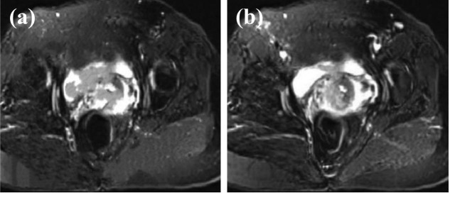
The aim of this research is to explore the application value of Deep residual network model (DRN) for deep learning-based multi-sequence magnetic resonance imaging (MRI) in the staging diagnosis of cervical cancer (CC). This research included 90 patients diagnosed with CC between August 2019 and May 2021 at the hospital. After undergoing MRI examination, the clinical staging and surgical pathological staging of patients were conducted. The research then evaluated the results of clinical staging and MRI staging to assess their diagnostic accuracy and correlation. In the staging diagnosis of CC, the feature enhancement layer was added to the DRN model, and the MRI imaging features of CC were used to enhance the image information. The precision, specificity, and sensitivity of the constructed model were analyzed, and then the accuracy of clinical diagnosis staging and MRI staging were compared. As the model constructed DRN in this research was compared with convolutional neural network (CNN) and the classic deep neural network visual geometry group (VGG), the precision was 67.7, 84.9, and 93.6%, respectively. The sensitivity was 70.4, 82.5, and 91.2%, while the specificity was 68.5, 83.8, and 92.2%, respectively. The precision, sensitivity, and specificity of the model were remarkably higher than those of CNN and VGG models ( < 0.05). As the clinical staging and MRI staging of CC were compared, the diagnostic accuracy of MRI was 100%, while that of clinical diagnosis was 83.7%, showing a significant difference between them ( < 0.05). Multi-sequence MRI under intelligent algorithm had a high diagnostic rate for CC staging, deserving a good clinical application value.

摘要

本研究旨在探讨深度残差网络模型(DRN)在基于深度学习的多序列磁共振成像(MRI)用于宫颈癌(CC)分期诊断中的应用价值。本研究纳入了2019年8月至2021年5月期间在该医院确诊为CC的90例患者。患者接受MRI检查后,进行了临床分期和手术病理分期。然后,该研究评估了临床分期和MRI分期的结果,以评估其诊断准确性和相关性。在CC的分期诊断中,在DRN模型中添加了特征增强层,并利用CC的MRI成像特征增强图像信息。分析了构建模型的精度、特异性和敏感性,然后比较了临床诊断分期和MRI分期的准确性。由于本研究构建的DRN模型与卷积神经网络(CNN)和经典深度神经网络视觉几何组(VGG)进行了比较,其精度分别为67.7%、84.9%和93.6%。敏感性分别为70.4%、82.5%和91.2%,而特异性分别为68.5%、83.8%和92.2%。该模型的精度、敏感性和特异性显著高于CNN和VGG模型(<0.05)。比较CC的临床分期和MRI分期,MRI的诊断准确性为100%,而临床诊断的诊断准确性为83.7%,两者之间存在显著差异(<0.05)。智能算法下的多序列MRI对CC分期具有较高的诊断率,具有良好的临床应用价值。

https://cdn.ncbi.nlm.nih.gov/pmc/blobs/20cb/11167709/b949ea85491c/j_biol-2022-0733-fig008.jpg
https://cdn.ncbi.nlm.nih.gov/pmc/blobs/20cb/11167709/a7bf09de184c/j_biol-2022-0733-fig001.jpg
https://cdn.ncbi.nlm.nih.gov/pmc/blobs/20cb/11167709/b4899f9a614a/j_biol-2022-0733-fig002.jpg
https://cdn.ncbi.nlm.nih.gov/pmc/blobs/20cb/11167709/854d3537003d/j_biol-2022-0733-fig003.jpg
https://cdn.ncbi.nlm.nih.gov/pmc/blobs/20cb/11167709/47446d85a530/j_biol-2022-0733-fig004.jpg
https://cdn.ncbi.nlm.nih.gov/pmc/blobs/20cb/11167709/d99c1a843280/j_biol-2022-0733-fig005.jpg
https://cdn.ncbi.nlm.nih.gov/pmc/blobs/20cb/11167709/38f8e5bf1631/j_biol-2022-0733-fig006.jpg
https://cdn.ncbi.nlm.nih.gov/pmc/blobs/20cb/11167709/444c7289e0f6/j_biol-2022-0733-fig007.jpg
https://cdn.ncbi.nlm.nih.gov/pmc/blobs/20cb/11167709/b949ea85491c/j_biol-2022-0733-fig008.jpg

相似文献

1
Application value of artificial intelligence algorithm-based magnetic resonance multi-sequence imaging in staging diagnosis of cervical cancer.基于人工智能算法的磁共振多序列成像在宫颈癌分期诊断中的应用价值
Open Life Sci. 2024 Jun 11;19(1):20220733. doi: 10.1515/biol-2022-0733. eCollection 2024.
2
Multimodal MRI Analysis of Cervical Cancer on the Basis of Artificial Intelligence Algorithm.基于人工智能算法的宫颈癌多模态 MRI 分析。
Contrast Media Mol Imaging. 2021 Nov 8;2021:1673490. doi: 10.1155/2021/1673490. eCollection 2021.
3
Magnetic Resonance Imaging Features on Deep Learning Algorithm for the Diagnosis of Nasopharyngeal Carcinoma.基于深度学习算法的磁共振成像特征在鼻咽癌诊断中的应用。
Contrast Media Mol Imaging. 2022 May 25;2022:3790269. doi: 10.1155/2022/3790269. eCollection 2022.
4
Diagnosis of Early Cervical Cancer with a Multimodal Magnetic Resonance Image under the Artificial Intelligence Algorithm.基于人工智能算法的多模态磁共振成像在早期宫颈癌诊断中的应用。
Contrast Media Mol Imaging. 2022 Mar 23;2022:6495309. doi: 10.1155/2022/6495309. eCollection 2022.
5
Image Features of Magnetic Resonance Imaging under the Deep Learning Algorithm in the Diagnosis and Nursing of Malignant Tumors.深度学习算法下磁共振成像的图像特征在恶性肿瘤诊断及护理中的应用
Contrast Media Mol Imaging. 2021 Aug 30;2021:1104611. doi: 10.1155/2021/1104611. eCollection 2021.
6
Application and Clinical Value of Machine Learning-Based Cervical Cancer Diagnosis and Prediction Model in Adjuvant Chemotherapy for Cervical Cancer: A Single-Center, Controlled, Non-Arbitrary Size Case-Control Study.基于机器学习的宫颈癌诊断和预测模型在宫颈癌辅助化疗中的应用及临床价值:一项单中心、对照、非随意大小病例对照研究。
Contrast Media Mol Imaging. 2022 Jun 15;2022:2432291. doi: 10.1155/2022/2432291. eCollection 2022.
7
Deep Learning Algorithm-Based MRI Image in the Diagnosis of Diabetic Macular Edema.基于深度学习算法的 MRI 图像在糖尿病性黄斑水肿诊断中的应用。
Contrast Media Mol Imaging. 2022 Mar 4;2022:1035619. doi: 10.1155/2022/1035619. eCollection 2022.
8
Artificial intelligence (AI) diagnostic tools: utilizing a convolutional neural network (CNN) to assess periodontal bone level radiographically-a retrospective study.人工智能(AI)诊断工具:利用卷积神经网络(CNN)评估牙周骨水平的放射影像——一项回顾性研究。
BMC Oral Health. 2022 Sep 13;22(1):399. doi: 10.1186/s12903-022-02436-3.
9
Evaluation and Monitoring of Endometrial Cancer Based on Magnetic Resonance Imaging Features of Deep Learning.基于深度学习的磁共振成像特征对子宫内膜癌的评估与监测。
Contrast Media Mol Imaging. 2022 Mar 18;2022:5198592. doi: 10.1155/2022/5198592. eCollection 2022.
10
[Application of convolutional neural network to risk evaluation of positive circumferential resection margin of rectal cancer by magnetic resonance imaging].卷积神经网络在直肠癌磁共振成像环周切缘阳性风险评估中的应用
Zhonghua Wei Chang Wai Ke Za Zhi. 2020 Jun 25;23(6):572-577. doi: 10.3760/cma.j.cn.441530-20191023-00460.

引用本文的文献

1
Artificial intelligence radiomics in the diagnosis, treatment, and prognosis of gynecological cancer: a literature review.人工智能影像组学在妇科癌症诊断、治疗及预后中的应用:文献综述
Transl Cancer Res. 2025 Apr 30;14(4):2508-2532. doi: 10.21037/tcr-2025-618. Epub 2025 Apr 27.

本文引用的文献

1
Application value of 18F-FDG PETCT imaging in the clinical initial diagnosis and follow-up of primary lesions of cervical cancer.18F-FDG PETCT成像在宫颈癌原发灶临床初步诊断及随访中的应用价值
Transl Cancer Res. 2020 Jun;9(6):4005-4010. doi: 10.21037/tcr-20-2085.
2
Semi-Supervised Support Vector Machine for Digital Twins Based Brain Image Fusion.基于数字孪生的脑图像融合的半监督支持向量机
Front Neurosci. 2021 Jul 9;15:705323. doi: 10.3389/fnins.2021.705323. eCollection 2021.
3
[The Value of MRI in Early Cervical Cancer Staging with Vaginal Filling of Ultrasound Gel].
[MRI在早期宫颈癌伴阴道超声凝胶填充分期中的价值]
Sichuan Da Xue Xue Bao Yi Xue Ban. 2021 May;52(3):523-527. doi: 10.12182/20210560307.
4
Is it enough only a single T2-weighted image in sagittal plane for diagnosis and staging uterine cervical cancer?仅矢状面的一张T2加权图像对于子宫颈癌的诊断和分期是否足够?
Eur J Radiol. 2021 Jun;139:109689. doi: 10.1016/j.ejrad.2021.109689. Epub 2021 Mar 31.
5
Diagnostic efficiency of whole-body F-FDG PET/MRI, MRI alone, and SUV and ADC values in staging of primary uterine cervical cancer.全身 F-FDG PET/MRI、单独 MRI 以及 SUV 和 ADC 值在原发性宫颈癌分期中的诊断效率。
Cancer Imaging. 2021 Jan 22;21(1):16. doi: 10.1186/s40644-020-00372-5.
6
2018 FIGO Staging Classification for Cervical Cancer: Added Benefits of Imaging.2018 FIGO 宫颈癌分期分类:影像学的附加益处。
Radiographics. 2020 Oct;40(6):1807-1822. doi: 10.1148/rg.2020200013. Epub 2020 Sep 18.
7
PET/MRI in Cervical Cancer: Associations Between Imaging Biomarkers and Tumor Stage, Disease Progression, and Overall Survival.正电子发射断层显像/磁共振成像在宫颈癌中的应用:影像生物标志物与肿瘤分期、疾病进展及总生存期的相关性
J Magn Reson Imaging. 2021 Jan;53(1):305-318. doi: 10.1002/jmri.27311. Epub 2020 Aug 14.
8
CNN-RNN Based Intelligent Recommendation for Online Medical Pre-Diagnosis Support.基于卷积神经网络-循环神经网络的在线医学预诊断支持智能推荐
IEEE/ACM Trans Comput Biol Bioinform. 2021 May-Jun;18(3):912-921. doi: 10.1109/TCBB.2020.2994780. Epub 2021 Jun 3.
9
Revised FIGO Staging for Cervical Cancer - A New Role for MRI.修订后的 FIGO 宫颈癌分期——MRI 的新作用。
Rofo. 2020 Oct;192(10):937-944. doi: 10.1055/a-1198-5729. Epub 2020 Jul 30.
10
Pre-treatment prediction of early response to chemoradiotherapy by quantitative analysis of baseline staging FDG-PET/CT and MRI in locally advanced cervical cancer.基于基线期 FDG-PET/CT 和 MRI 定量分析预测局部晚期宫颈癌放化疗早期应答。
Acta Radiol. 2021 Jul;62(7):940-948. doi: 10.1177/0284185120943046. Epub 2020 Jul 28.