Department of Radiology, Turku University Hospital and University of Turku, PO Box 52, 20521, Turku, Finland.
Department of Radiology, Brigham and Women's Hospital, 75 Francis St, Boston, MA, 02115, USA.
Cancer Imaging. 2021 Jan 22;21(1):16. doi: 10.1186/s40644-020-00372-5.
The use of PET/MRI for gynecological cancers is emerging. The purpose of this study was to assess the additional diagnostic value of PET over MRI alone in local and whole-body staging of cervical cancer, and to evaluate the benefit of standardized uptake value (SUV) and apparent diffusion coefficient (ADC) in staging.
Patients with histopathologically-proven cervical cancer and whole-body F-FDG PET/MRI obtained before definitive treatment were retrospectively registered. Local tumor spread, nodal involvement, and distant metastases were evaluated using PET/MRI or MRI dataset alone. Histopathology or clinical consensus with follow-up imaging were used as reference standard. Tumor SUVmax and ADC were measured and SUVmax/ADC ratio calculated. Area under the curve (AUC) was determined to predict diagnostic performance and Mann-Whitney U test was applied for group comparisons.
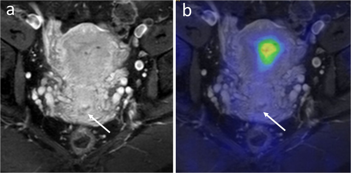
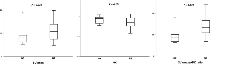
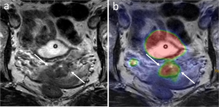
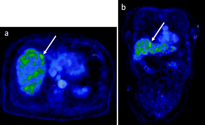
In total, 33 patients who underwent surgery (n = 23) or first-line chemoradiation (n = 10) were included. PET/MRI resulted in higher AUC compared with MRI alone in detecting parametrial (0.89 versus 0.73), vaginal (0.85 versus 0.74), and deep cervical stromal invasion (0.96 versus 0.74), respectively. PET/MRI had higher diagnostic confidence than MRI in identifying patients with radical cone biopsy and no residual at hysterectomy (sensitivity 89% versus 44%). PET/MRI and MRI showed equal AUC for pelvic nodal staging (both 0.73), whereas AUC for distant metastases was higher using PET/MRI (0.80 versus 0.67). Tumor SUVmax/ADC ratio, but not SUVmax or ADC alone, was significantly higher in the presence of metastatic pelvic lymph nodes (P < 0.05).
PET/MRI shows higher accuracy than MRI alone for determining local tumor spread and distant metastasis emphasizing the added value of PET over MRI alone in staging of cervical cancer. Tumor SUVmax/ADC ratio may predict pelvic nodal involvement.
PET/MRI 在妇科癌症中的应用正在兴起。本研究旨在评估 PET 相对于单独 MRI 在宫颈癌局部和全身分期中的额外诊断价值,并评估标准摄取值(SUV)和表观扩散系数(ADC)在分期中的益处。
回顾性登记了经组织病理学证实的宫颈癌患者和全身 F-FDG PET/MRI 检查结果。使用 PET/MRI 或 MRI 数据集单独评估局部肿瘤扩散、淋巴结受累和远处转移。将组织病理学或临床共识与随访影像学检查作为参考标准。测量肿瘤 SUVmax 和 ADC,并计算 SUVmax/ADC 比值。确定曲线下面积(AUC)以预测诊断性能,并应用 Mann-Whitney U 检验进行组间比较。
共纳入 33 例接受手术(n=23)或一线放化疗(n=10)的患者。与单独 MRI 相比,PET/MRI 在检测宫旁(0.89 对 0.73)、阴道(0.85 对 0.74)和深部宫颈间质浸润(0.96 对 0.74)方面具有更高的 AUC。与 MRI 相比,PET/MRI 在识别接受根治性宫颈锥切术且子宫切除术后无残留的患者时具有更高的诊断信心(敏感性 89%对 44%)。PET/MRI 和 MRI 对盆腔淋巴结分期的 AUC 相等(均为 0.73),而 PET/MRI 对远处转移的 AUC 更高(0.80 对 0.67)。存在转移性盆腔淋巴结时,肿瘤 SUVmax/ADC 比值显著升高(P<0.05),而 SUVmax 或 ADC 单独升高则不显著。
与单独 MRI 相比,PET/MRI 对确定局部肿瘤扩散和远处转移具有更高的准确性,强调了 PET 相对于单独 MRI 在宫颈癌分期中的附加价值。肿瘤 SUVmax/ADC 比值可能预测盆腔淋巴结受累。